This cadaveric dissection provides a high-fidelity view of the carotid triangle, a vital anatomical region within the neck. By examining the transition from the common carotid artery to its specialized terminal branches, healthcare professionals can better understand the vascular supply to the head and the critical nerves and muscles that facilitate speech and swallowing.
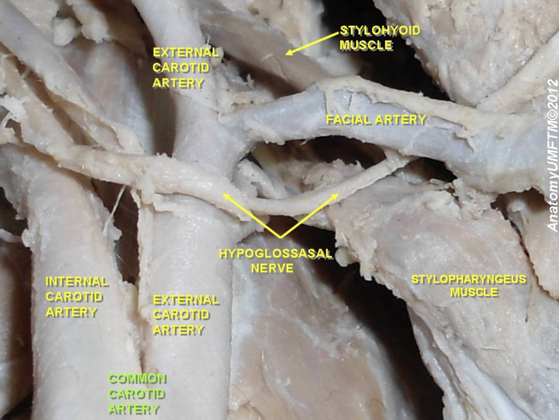
EXTERNAL CAROTID ARTERY: This major arterial branch is responsible for providing oxygenated blood to the majority of structures in the face, scalp, and neck. Unlike the internal carotid artery, the external carotid gives off eight distinct branches in the neck, including the superior thyroid and lingual arteries.
STYLOHYOID MUSCLE: A slender muscle belonging to the suprahyoid group, it originates from the styloid process of the temporal bone. Its primary physiological function is to elevate and retract the hyoid bone during the swallowing process and during vocalization.
FACIAL ARTERY: This is a significant branch of the external carotid artery that travels deep to the submandibular gland before crossing the mandible. It provides the essential blood supply for the muscles of facial expression and follows a characteristically tortuous path.
HYPOGLOSSASAL NERVE: This is the twelfth cranial nerve (CN XII), which provides motor control to nearly all the intrinsic and extrinsic muscles of the tongue. In this dissection, it is seen crossing the carotid vessels superficially, serving as a critical surgical landmark during carotid endarterectomy procedures.
STYLOPHARYNGEUS MUSCLE: A long, thin muscle that descends from the styloid process to the pharyngeal wall and thyroid cartilage. It is the only muscle innervated by the glossopharyngeal nerve (CN IX) and helps elevate the larynx and pharynx during deglutition.
INTERNAL CAROTID ARTERY: This vessel is the primary conduit for oxygenated blood to the brain and the orbital structures. It is unique among cervical arteries as it gives off no branches until it enters the skull through the carotid canal.
COMMON CAROTID ARTERY: The main arterial trunk that ascends within the carotid sheath along the neck. It bifurcates into the internal and external branches typically at the level of the superior border of the thyroid cartilage.
Functional Significance of the Carotid Triangle
The area depicted in the image is known as the carotid triangle, a subsection of the anterior triangle of the neck defined by the sternocleidomastoid, the superior belly of the omohyoid, and the posterior belly of the digastric muscle. This space is clinically significant because it houses the carotid bifurcation, where the body’s blood pressure and chemical sensors are located. The carotid sinus, found at the base of the internal carotid artery, contains baroreceptors that detect changes in arterial pressure to maintain hemodynamic stability through autonomic feedback loops.
Within this region, the coordination between vascular supply and neural control is paramount. The presence of the hypoglossal nerve (CN XII) crossing the arteries illustrates how closely the nervous system is integrated with the circulatory system in the neck. Damage to these nerves during surgical interventions can lead to significant functional deficits, such as difficulties in speech (dysarthria) or swallowing (dysphagia).
Key features of the carotid system include:
- The carotid body, a chemoreceptor located at the bifurcation that monitors oxygen levels in the blood.
- The carotid sheath, a fascial layer that encloses the common carotid artery, internal jugular vein, and vagus nerve.
- The precise branching pattern of the external carotid artery, which ensures constant perfusion to the facial tissues.
- The relationship with the suprahyoid muscles, which facilitate the mechanical aspects of eating and breathing.
Physiological Coordination in the Neck
Physiologically, the carotid system is designed to prioritize cerebral perfusion above almost all other bodily needs. The internal carotid artery travels directly into the Circle of Willis, ensuring that the brain receives a steady stream of oxygen and glucose. Any obstruction in this pathway, such as atherosclerotic plaque, can severely compromise neurological function and increase the risk of ischemic events.
Furthermore, the muscles labeled in the dissection, such as the stylohyoid and stylopharyngeus, demonstrate the complex mechanical labor required for daily survival. These muscles work in sync with the larynx and pharynx to ensure that food is directed into the esophagus while protecting the airway. The blood supply from the facial artery and other external carotid branches supports the high metabolic demand of these frequently used muscular structures.
A thorough understanding of these anatomical relationships is indispensable for medical students, surgeons, and clinicians. By studying cadaveric images, one can appreciate the three-dimensional depth and slight variations that occur in human anatomy, moving beyond simplified textbook diagrams. This knowledge ensures safer surgical approaches and more accurate clinical diagnoses in patients presenting with cervical or vascular pathologies.
Mastering the intricate details of the carotid bifurcation and its neighboring structures is a cornerstone of medical education. The interplay between the XII cranial nerve and the major vascular trunks highlights the vulnerability and complexity of the human neck. Through continued study of these specimens, healthcare providers can refine their diagnostic skills and provide more effective care for complex neurovascular conditions.

