The human cardiovascular system is a marvel of biological engineering, ensuring oxygen and nutrients reach every cell while removing waste products. This diagram illustrates the dual circuits—pulmonary and systemic—that work in tandem to maintain life, with clear annotations highlighting the flow and oxygenation of blood through key organs and vessels.
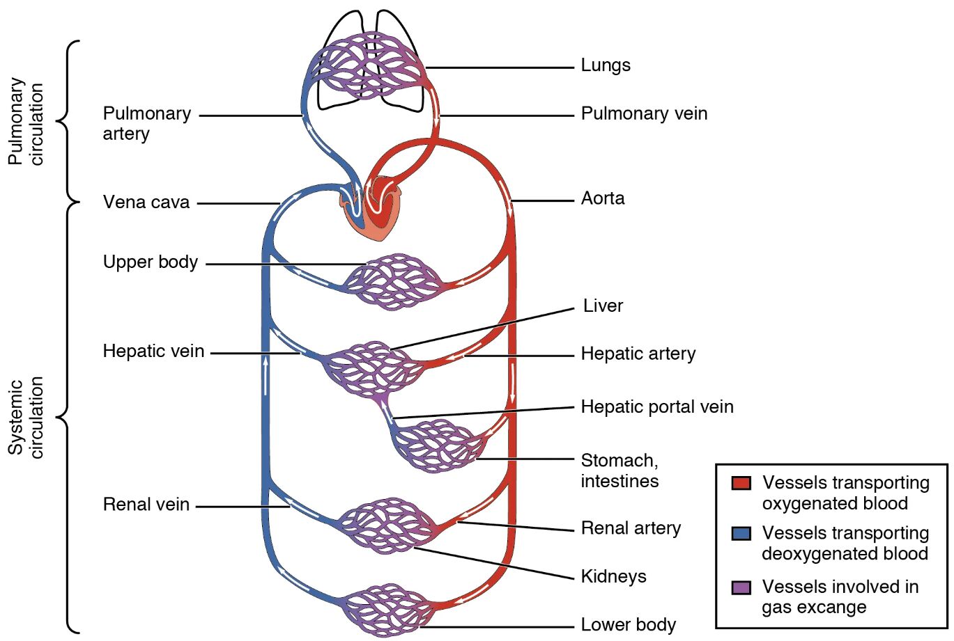
Pulmonary artery The pulmonary artery carries deoxygenated blood from the right ventricle of the heart to the lungs for oxygenation. It is a critical component of the pulmonary circulation, branching into smaller vessels to facilitate gas exchange in the lung capillaries.
Vena cava The vena cava, comprising the superior and inferior veins, returns deoxygenated blood from the upper and lower body to the right atrium. This large vein plays a vital role in the systemic circulation, ensuring continuous blood flow back to the heart for reoxygenation.
Upper body The upper body represents the region above the diaphragm, including the head, neck, and arms, where deoxygenated blood is collected by the superior vena cava. It receives oxygenated blood from the aorta, supporting metabolic activities in these areas.
Hepatic vein The hepatic vein drains deoxygenated blood from the liver into the inferior vena cava, completing the hepatic portal circulation loop. It transports nutrient-rich blood processed by the liver back to the heart for recirculation.
Renal vein The renal vein carries filtered blood from the kidneys to the inferior vena cava after waste removal and fluid balance adjustment. It ensures that cleansed blood re-enters the systemic circulation efficiently.
Lungs The lungs are the primary sites for gas exchange, where deoxygenated blood becomes oxygenated through the alveoli. They receive blood via the pulmonary artery and return it to the heart via the pulmonary vein.
Pulmonary vein The pulmonary vein transports oxygenated blood from the lungs back to the left atrium of the heart, marking the end of the pulmonary circuit. This vessel is unique as it carries oxygenated blood, unlike most veins.
Aorta The aorta is the largest artery, carrying oxygenated blood from the left ventricle to the systemic circulation, distributing it to the body’s organs and tissues. It branches into major arteries, ensuring widespread oxygen delivery.
Liver The liver receives blood via the hepatic artery and hepatic portal vein, processing nutrients and detoxifying substances. It plays a key role in metabolism, influencing blood composition before it returns to the heart.
Hepatic artery The hepatic artery supplies oxygenated blood directly from the aorta to the liver, supporting its metabolic and synthetic functions. This vessel ensures the liver receives adequate oxygen for its diverse physiological roles.
Hepatic portal vein The hepatic portal vein carries nutrient-rich, deoxygenated blood from the stomach and intestines to the liver for processing. It facilitates the liver’s role in regulating blood glucose and storing vitamins.
Stomach, intestines The stomach and intestines absorb nutrients and release them into the hepatic portal vein, contributing to the digestive system’s integration with circulation. These organs rely on systemic blood supply for their metabolic needs.
Renal artery The renal artery delivers oxygenated blood from the aorta to the kidneys, supporting filtration and waste excretion. It ensures the kidneys maintain homeostasis through efficient blood flow.
Kidneys The kidneys filter blood to remove waste, regulate electrolytes, and produce urine, receiving blood via the renal artery. They return cleansed blood to the systemic circulation through the renal vein.
Lower body The lower body, including the legs and pelvic region, receives oxygenated blood from the aorta and returns deoxygenated blood via the inferior vena cava. This region depends on robust circulation for movement and tissue health.
The Role of Pulmonary Circulation
The pulmonary circulation is essential for oxygenating blood, a process that sustains life across all bodily systems. This circuit begins and ends with the heart, ensuring efficient gas exchange in the lungs.
- Blood leaves the right ventricle through the pulmonary artery, traveling to the lungs.
- Within the lungs, carbon dioxide is expelled, and oxygen is absorbed into the bloodstream.
- The pulmonary vein then carries this oxygenated blood back to the left atrium.
- This cycle operates continuously, adapting to the body’s oxygen demands.
- Hormones like erythropoietin, produced in the kidneys, influence red blood cell production to support this process.
The Systemic Circulation Pathway
Systemic circulation delivers oxygenated blood to the body’s tissues, a critical function for maintaining cellular health. This circuit starts at the left ventricle, branching out to nourish every organ.
- The aorta distributes oxygen-rich blood to arteries like the hepatic and renal arteries.
- Deoxygenated blood returns via veins such as the vena cava and renal vein.
- The liver and kidneys process this blood, removing waste and regulating substances.
- The lower and upper body receive tailored blood flow based on metabolic needs.
- Thyroid hormones like T3 and T4 can enhance cardiac output to support this circulation.
Anatomical Highlights of Key Vessels
The diagram’s vessels are color-coded to reflect their oxygen content, providing a clear visual guide to blood flow. Each vessel serves a specific role in the dual circulatory system.
- The pulmonary artery is depicted in blue, indicating its deoxygenated load.
- Red hues mark the aorta and hepatic artery, signifying oxygenated blood.
- Purple areas in the lungs highlight vessels involved in gas exchange.
- The vena cava and renal vein are blue, showing their deoxygenated return flow.
- These color distinctions aid in understanding circulatory dynamics.
Physiological Functions of Major Organs
Organs like the lungs, liver, and kidneys are integral to circulation, each performing specialized tasks. Their interactions with the cardiovascular system ensure homeostasis.
- The lungs oxygenate blood and remove carbon dioxide, driven by respiratory mechanics.
- The liver metabolizes nutrients and detoxifies blood, supported by dual blood supply.
- Kidneys filter waste and regulate blood pressure through renin-angiotensin mechanisms.
- The stomach and intestines absorb nutrients, feeding into the hepatic portal system.
- The heart acts as the pump, coordinating both circuits seamlessly.
Comparative Circulation Across Species
Human circulation shares similarities with other mammals, though adaptations vary. Studying these differences enhances our understanding of cardiovascular evolution.
- Mammals like mice have similar pulmonary and systemic loops, scaled to size.
- Fish rely on a single-circuit system, contrasting with the human dual setup.
- Birds exhibit high-efficiency circulation for flight, with unique vessel adaptations.
- Reptiles show partial separation, offering insights into evolutionary transitions.
- These comparisons inform research into congenital circulatory anomalies.
Clinical Insights and Monitoring
Understanding this diagram aids in diagnosing circulatory issues through imaging and tests. Early detection can prevent complications in related systems.
- Echocardiograms assess heart and vessel function in pulmonary circulation.
- Blood tests measure liver and kidney efficiency in systemic circulation.
- Doppler ultrasound tracks blood flow in the renal and hepatic arteries.
- Abnormalities in the vena cava may indicate clotting or heart strain.
- Regular monitoring supports timely interventions for circulatory health.
Future Advances in Cardiovascular Research
Ongoing studies aim to improve circulatory health using this anatomical framework. Innovations promise better treatments for vascular conditions.
- Stem cell therapy targets vessel regeneration in the lungs and kidneys.
- 3D modeling of the aorta aids in surgical planning for aneurysms.
- Gene therapy explores enhancing hepatic portal vein function.
- Wearable devices monitor real-time blood flow in the upper and lower body.
- Collaborative efforts integrate these advances for patient care.
This detailed diagram of cardiovascular circulation offers a window into the body’s life-sustaining network, where every vessel and organ plays a coordinated role. Exploring these pathways deepens appreciation for the heart’s relentless work and inspires continued research to enhance human health.

