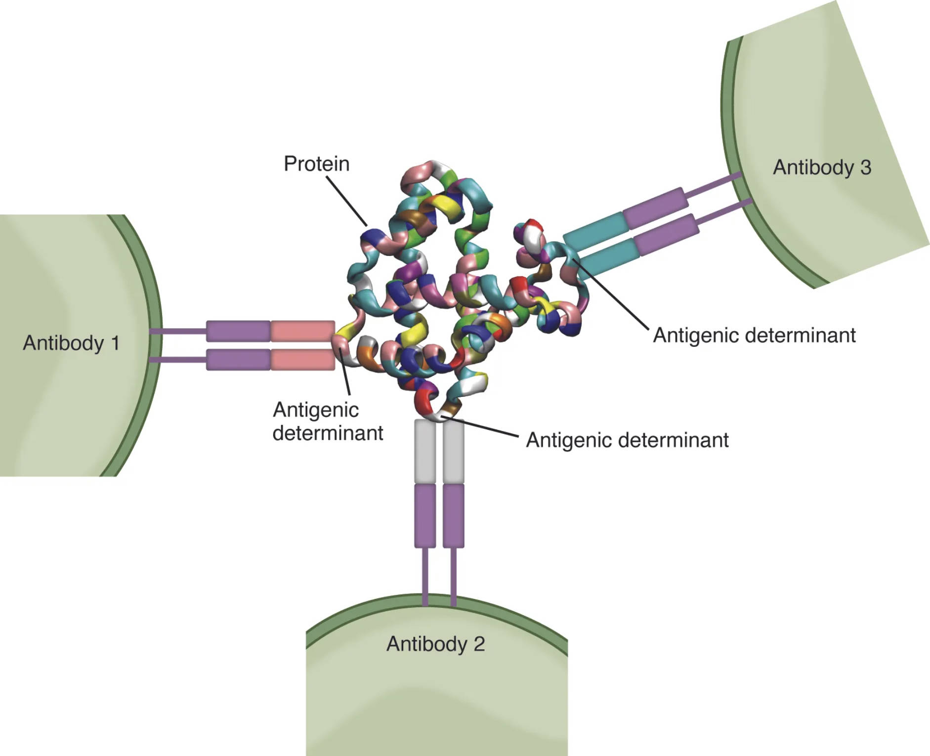
Antigenic determinants, or epitopes, are specific regions on an antigen that the immune system recognizes and targets, playing a pivotal role in immune defense. A typical protein antigen features multiple antigenic determinants, allowing T cells with different specificities to bind and initiate a tailored response, as illustrated in this detailed image. This visual representation highlights how these determinants drive the diversity and precision of immune reactions, offering a deeper look into the mechanisms of immunity.
Labeled Components of Antigenic Determinants
Antigen: This foreign molecule, often a protein, triggers an immune response when introduced into the body. It contains multiple antigenic determinants that are recognized by T cells and antibodies.
Antigenic determinant (epitope 1): This specific site on the antigen is recognized by a particular T cell receptor or antibody. Its unique structure allows for targeted immune binding and activation.
Antigenic determinant (epitope 2): Another distinct region on the same antigen, this epitope is bound by a different T cell with unique specificity. It contributes to the antigen’s overall immunogenicity.
Antigenic determinant (epitope 3): This third epitope on the antigen is recognized by yet another T cell, showcasing the antigen’s ability to engage multiple immune cells. Its presence enhances the breadth of the immune response.
T cell receptor (TCR 1): This receptor on a T cell binds specifically to epitope 1, initiating an immune response. It is tailored to recognize a particular antigenic determinant presented by MHC molecules.
T cell receptor (TCR 2): Designed to bind epitope 2, this TCR on a different T cell activates a distinct immune pathway. It highlights the diversity of T cell recognition.
T cell receptor (TCR 3): This receptor targets epitope 3, engaging a third T cell in the response. Its specificity underscores the complexity of antigen recognition.
T cell (T cell 1): This immune cell, equipped with TCR 1, responds to epitope 1 by proliferating or releasing cytokines. It plays a key role in coordinating the immune attack.
T cell (T cell 2): Carrying TCR 2, this T cell targets epitope 2, contributing to a multifaceted immune strategy. It enhances the overall effectiveness against the antigen.
T cell (T cell 3): With TCR 3, this T cell binds epitope 3, adding another layer of immune specificity. Its activation ensures comprehensive antigen clearance.
MHC molecule: This major histocompatibility complex molecule presents the antigenic determinant to the T cell receptor. It is essential for T cell recognition of processed antigens.
Anatomical Context of Antigenic Determinants
Antigenic determinants are integral to how the immune system identifies and combats pathogens.
- The antigen serves as the target, with multiple epitopes exposed for recognition.
- Epitope 1, 2, and 3 each attract specific T cell receptors, enabling diverse responses.
- T cell receptors 1, 2, and 3 are uniquely shaped to match their respective epitopes.
- T cells 1, 2, and 3 activate upon binding, initiating tailored immune actions.
- MHC molecules on antigen-presenting cells display epitopes to T cells.
- This multi-determinant recognition enhances the immune system’s adaptability.
This illustration emphasizes the antigen’s role as a multifaceted target.
Physiological Role in Immune Recognition
Antigenic determinants drive the specificity and versatility of immune responses.
- The antigen’s multiple epitopes allow simultaneous engagement by different T cells.
- T cell receptors bind with high affinity to their specific epitopes, triggering activation.
- T cells proliferate or differentiate into effector cells upon recognition.
- MHC molecules ensure proper presentation, linking innate and adaptive immunity.
- This process generates memory T cells for future encounters with the antigen.
- The diversity of epitopes supports broad protection against pathogens.
This mechanism is key to the immune system’s ability to adapt.
Molecular Structure of Antigenic Determinants
The structure of antigenic determinants determines their immunogenicity and recognition.
- Epitopes are typically short peptide sequences or conformational shapes on the antigen.
- T cell receptors recognize epitopes in the context of MHC molecules.
- The antigen’s three-dimensional folding exposes multiple binding sites.
- MHC molecules process and present epitopes, altering their accessibility.
- T cell activation depends on the precise fit between TCR and epitope.
- Variability in epitope structure influences the strength of the immune response.
This structural diversity is critical for effective immunity.
Clinical Relevance of Antigenic Determinants
Understanding antigenic determinants aids in managing immune-related conditions.
- Altered epitope recognition can lead to autoimmune diseases, where T cells attack self-antigens.
- Vaccine design targets specific epitopes to elicit protective T cell responses.
- MHC molecule variations affect epitope presentation, impacting transplant success.
- Multiple epitopes on a pathogen can complicate antiviral drug development.
- T cell receptor diversity is studied in immunodeficiencies affecting antigen recognition.
- Cancer immunotherapy leverages epitope-specific T cells to target tumors.
This knowledge supports innovative treatments and diagnostics.
Developmental and Adaptive Features
Antigenic determinants shape the immune system’s evolution and memory.
- T cell receptors develop diversity through V(D)J recombination in the thymus.
- Exposure to antigens refines T cell specificity, enhancing epitope recognition.
- MHC molecules adapt to present a wide range of epitopes over time.
- T cells gain memory, improving responses to previously encountered epitopes.
- The antigen’s multiple determinants drive clonal expansion of T cells.
- This adaptability ensures long-term immunity against diverse threats.
This process highlights the immune system’s learning capacity.
Antigenic determinants, as depicted in this illustration, are the cornerstone of immune specificity, with a single antigen engaging multiple T cells through distinct epitopes. This cooperative recognition, facilitated by MHC molecules and T cell receptors, underscores the body’s remarkable ability to mount a tailored defense, making it a fascinating area of study for those interested in immunology and health.

