Neurons, the fundamental building blocks of the nervous system, exhibit diverse shapes that reflect their specific roles in processing and conveying information throughout the body. This diagram classifies neurons based on their morphology into unipolar, bipolar, and multipolar types, each adapted for distinct functional demands in sensory perception, signal relay, or integration. Such classification underscores the efficiency of neuronal design, where the number and arrangement of processes—extensions like axons and dendrites—optimize communication pathways in both the central and peripheral nervous systems.
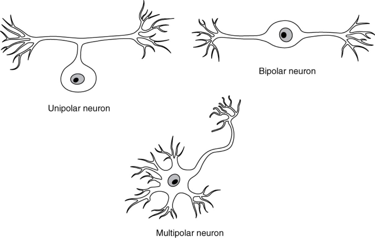
Labeled Types of Neurons
Unipolar Neuron
The **unipolar neuron features a single process extending from the cell body, which branches into a peripheral process acting as a dendrite and a central process functioning as an axon. This structure is commonly found in sensory neurons of the peripheral nervous system, allowing efficient transmission of touch or pain signals from the skin to the spinal cord.
Bipolar Neuron
Bipolar neurons possess two distinct processes: one axon and one dendrite, each emerging from opposite ends of the cell body that remains relatively simple and rounded. These neurons are prevalent in specialized sensory systems, such as the retina for visual processing or in the inner ear for smell detection, where they relay information from sensory receptors to higher neural circuits with minimal branching.
Multipolar Neuron
Multipolar neurons are defined by one axon and multiple dendrites branching from the cell body, enabling extensive connectivity for complex signal integration. This configuration dominates in the central nervous system, particularly in motor neurons that control muscle movement and interneurons that facilitate intricate brain computations.
In-Depth Anatomy of Neuron Types
Neuron shapes are determined during development by genetic cues and environmental factors, influencing how they integrate into neural networks. Each type’s anatomy supports unique physiological demands, from rapid sensory input to multifaceted processing.
- Unipolar neurons, often pseudounipolar in mammals, have the single process splitting near the cell body into axonal and dendritic segments, minimizing the distance for action potentials to travel from peripheral receptors.
- In bipolar neurons, the dendrite typically interfaces with sensory cells, such as photoreceptors in the retina, while the axon projects to ganglion cells, forming a streamlined pathway for initial signal amplification.
- Multipolar neurons exhibit varied dendritic trees, with spines increasing synaptic sites, as seen in hippocampal neurons involved in learning.
- The cell body in all types contains Nissl substance for protein synthesis, but in multipolar forms, it’s more pronounced to sustain extensive branching.
- Axonal lengths differ; unipolar axons can extend over long distances in sensory pathways, whereas bipolar ones are shorter for local relay.
Physiological Roles and Adaptations
The morphology of neurons directly impacts their electrical properties and integration within circuits. Adaptations in shape enhance efficiency in signal conduction and synaptic interactions.
- Unipolar neurons facilitate fast, unidirectional flow, with the peripheral branch detecting stimuli via mechanoreceptors or nociceptors, and the central branch synapsing in dorsal root ganglia.
- Bipolar neurons excel in graded potential transmission, as in cochlear hair cells where they convert sound vibrations into electrical signals without full action potentials.
- Multipolar neurons support summation of multiple inputs, with dendrites integrating excitatory postsynaptic potentials (EPSPs) from thousands of synapses to reach firing thresholds.
- In terms of membrane properties, all types maintain a resting potential via Na+/K+ pumps, but multipolar dendrites often have voltage-gated channels for active propagation.
- Myelination varies; unipolar peripheral processes may be heavily myelinated by Schwann cells for speed in afferent nerves.
Neuron Types in the Peripheral and Central Nervous Systems
Distribution across the body highlights functional specialization. Peripheral neurons handle input and output, while central ones manage higher-order tasks.
- Unipolar neurons predominate in the peripheral nervous system (PNS), such as in dorsal root ganglia for somatosensory information and cranial nerves for facial sensations.
- Bipolar neurons are rarer, primarily in special senses: retinal bipolar cells bipolar cells modulate visual signals between photoreceptors and ganglion cells, aiding contrast detection.
- Multipolar neurons form the core of the central nervous system (CNS), like pyramidal cells in the cerebral cortex for cognition or alpha motor neurons in the spinal cord for muscle innervation.
- Transitions occur; some PNS neurons project into CNS, ensuring continuity in reflex arcs.
- Evolutionary, bipolar forms may represent primitive states, with multipolar evolving for advanced vertebrate brains.
Developmental and Evolutionary Aspects of Neurons
Neuron shapes emerge from progenitor cells through differentiation processes. Evolutionary trends show increasing complexity for behavioral sophistication.
- During neurogenesis, polarity is established by cytoskeletal rearrangements involving Rho GTPases, guiding process outgrowth in bipolar configurations.
- Unipolar morphology in invertebrates like nematodes contrasts with mammalian pseudounipolar adaptations for ganglion clustering.
- Bipolar neurons in olfactory epithelium regenerate throughout life, supported by basal stem cells, unlike most CNS neurons.
- Multipolar development includes dendritic pruning, refined by activity-dependent mechanisms like long-term potentiation (LTP).
- Genetic factors, such as Neurogenin transcription factors, dictate type-specific differentiation in neural crests.
Research Techniques for Studying Neuron Morphology
Modern methods provide detailed visualizations and manipulations of neuron shapes.
- Golgi staining selectively labels entire neurons, revealing dendritic arbors in multipolar cells for quantitative analysis.
- Patch-clamp recordings in bipolar neurons measure ionic currents, elucidating roles in sensory transduction.
- Two-photon microscopy tracks live unipolar process dynamics in peripheral tissues.
- CRISPR editing alters genes for shape, studying impacts on circuit function.
- Computational models simulate signal flow in different morphologies using NEURON software.
Pathologies Associated with Neuron Types
Structural anomalies in neuron shapes contribute to various disorders.
- Damage to unipolar sensory neurons in diabetic neuropathy leads to loss of sensation due to axonal degeneration.
- Bipolar cell dysfunction in congenital stationary night blindness impairs rod-cone signaling in the retina.
- Multipolar neuron loss in amyotrophic lateral sclerosis (ALS) affects motor control, with glutamate toxicity causing dendritic retraction.
- Neurodevelopmental disorders like autism may involve altered dendritic branching in multipolar cortical neurons.
- Regeneration challenges differ; PNS unipolar axons regrow more readily than CNS multipolar ones due to inhibitory myelin.
In conclusion, the classification of neurons into unipolar, bipolar, and multipolar types illustrates the elegant diversity tailored to neural functions, from basic sensory relay to sophisticated integration. Appreciating these structures enhances comprehension of nervous system operations and informs approaches to treating related pathologies, driving continued progress in neuroscience.

