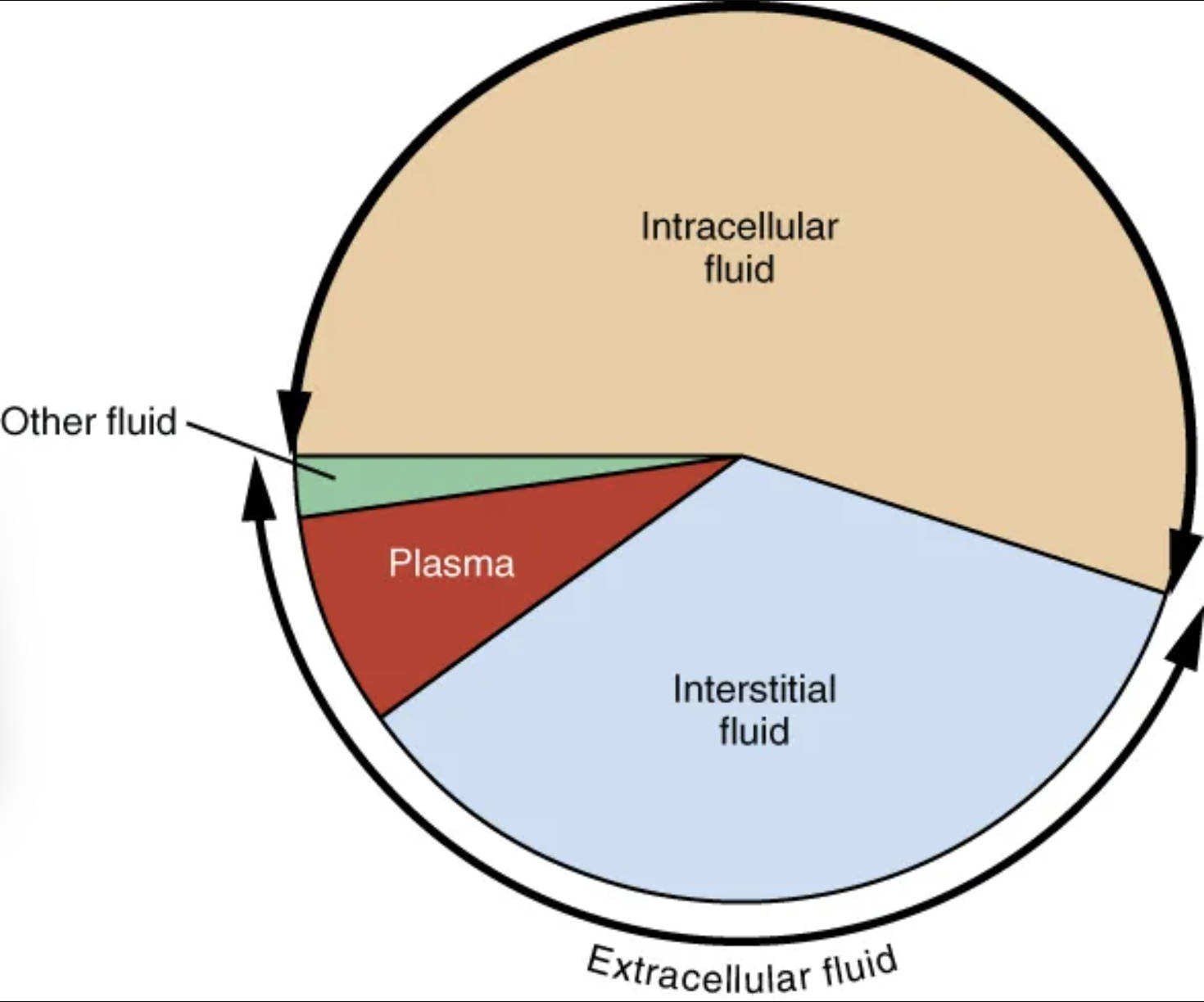
The human body is remarkably adept at maintaining its internal environment, a critical aspect of which is the precise distribution of water. This pie graph visually represents how the total body fluid is partitioned into distinct compartments: intracellular fluid, interstitial fluid, plasma, and other fluids. Understanding these proportions is fundamental for grasping concepts related to fluid balance, electrolyte homeostasis, and the physiological responses to various health conditions. This visual aid simplifies the complex world of fluid dynamics, offering a foundational understanding of where the body’s essential water resides.

Intracellular fluid: Intracellular fluid (ICF) is the fluid contained within the cells.
The intracellular fluid (ICF) constitutes the largest single fluid compartment in the body, accounting for approximately two-thirds of the total body water. This fluid is vital for numerous cellular processes, serving as the medium for metabolic reactions and maintaining the cell’s structural integrity. The unique composition of the ICF is tightly regulated, differing significantly from the fluid outside the cells, and is crucial for optimal cell function.
Other fluid: This category includes transcellular fluids, such as cerebrospinal fluid, synovial fluid, and intraocular fluid.
The “Other fluid” category represents a relatively small but functionally significant portion of the total body fluid. These are specialized fluids found in various body cavities and spaces, including cerebrospinal fluid cushioning the brain and spinal cord, synovial fluid lubricating joints, and intraocular fluid maintaining eye pressure. Despite their small volume, these fluids play critical roles in protection, lubrication, and nutrient transport in specific areas.
Plasma: Plasma is the liquid component of blood, the intravascular fluid.
Plasma makes up approximately 20-25% of the extracellular fluid (ECF) and is the fluid medium in which blood cells are suspended. It is essential for the transport of nutrients, hormones, oxygen, and metabolic waste products throughout the circulatory system. Maintaining adequate plasma volume is crucial for blood pressure regulation and overall cardiovascular function.
Interstitial fluid: Interstitial fluid (IF) is the fluid that surrounds the cells, not including blood cells.
The interstitial fluid (IF) represents the second largest fluid compartment in the body and the primary component of the extracellular fluid (ECF), typically accounting for about 75-80% of the ECF. It acts as the immediate environment for cells, facilitating the exchange of substances between the blood plasma and the intracellular fluid. This fluid is crucial for delivering nutrients and oxygen to cells while removing waste products.
Extracellular fluid: Extracellular fluid (ECF) encompasses all fluid outside the cells, including plasma, interstitial fluid, and transcellular fluids.
The extracellular fluid (ECF) is the fluid found outside the cells, acting as the body’s internal environment. It is further subdivided into interstitial fluid, plasma, and the “other fluids” (transcellular fluids). The ECF is responsible for transporting substances to and from cells, maintaining the stability of the cellular environment, and playing a vital role in the body’s homeostatic mechanisms.
The graphical representation clearly illustrates that the vast majority of the body’s water is held within the cells themselves, emphasizing the cellular dependence on water for all life processes. The remaining portion, the extracellular fluid (ECF), serves as the dynamic bridge between the external environment and the internal cellular milieu. This distribution is not arbitrary; it reflects the body’s sophisticated mechanisms for nutrient delivery, waste removal, and overall physiological stability.
The continuous movement of water and solutes between these compartments is driven by osmotic and hydrostatic pressures, ensuring a delicate balance known as fluid homeostasis. Any disruption to this balance, whether due to illness, injury, or inadequate fluid intake, can have significant physiological consequences. For instance:
- Dehydration primarily impacts the extracellular fluid first, leading to reduced blood volume.
- Certain conditions can cause fluid shifts, such as edema, where excessive fluid accumulates in the interstitial space.
- Electrolyte imbalances often manifest through altered fluid distribution across these compartments.
Understanding these fluid compartments is paramount for comprehending how the body regulates its internal environment and responds to various challenges. It provides a fundamental framework for assessing hydration status, diagnosing fluid imbalances, and implementing appropriate therapeutic interventions in a clinical setting. The interplay between these compartments is a testament to the body’s remarkable capacity for self-regulation.
In summary, this pie graph serves as an excellent visual aid for appreciating the intricate partitioning of total body fluid. The dominance of intracellular fluid, alongside the crucial roles of interstitial fluid and plasma within the extracellular compartment, underscores the complexity and precision of fluid regulation. A comprehensive understanding of these fluid dynamics is not only essential for physiological knowledge but also forms a cornerstone for effective clinical assessment and management of fluid and electrolyte disorders.

