The thoracic region of the spine demonstrates complex ligamentous attachments that are crucial for maintaining stability while enabling respiratory movement. These specialized ligamentous structures coordinate with osseous elements to create a sophisticated system that balances mobility with structural integrity. Understanding these relationships is essential for medical professionals involved in treating thoracic spine conditions.
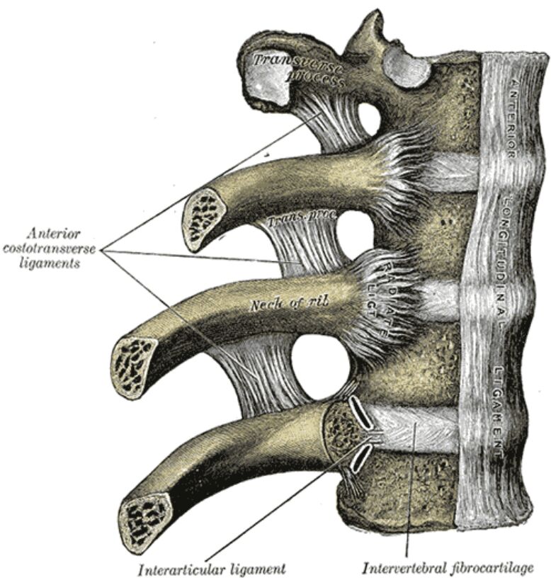
Transverse Process The transverse process extends laterally from the vertebral arch. This bony projection serves as an attachment point for multiple ligaments and muscles while providing articulation for the ribs in thoracic vertebrae.
Anterior costotransverse ligaments The anterior costotransverse ligaments connect the anterior aspect of the transverse process to the neck of the corresponding rib. These ligaments provide essential stability during respiratory movements while maintaining proper rib alignment.
Neck of rib The neck of the rib represents the portion between the head and tubercle. This region serves as an attachment point for multiple ligaments and demonstrates specialized architectural features for force transmission.
Interarticular ligament The interarticular ligament connects the crest of the rib neck to the intervertebral disc. This ligament provides additional stability to the costovertebral joint while allowing controlled movement during respiration.
Intervertebral fibrocartilage The intervertebral fibrocartilage forms the disc between vertebral bodies. Its complex structure enables both stability and mobility while providing crucial shock absorption for the spine.
Thoracic Ligament Architecture and Function
The thoracic spine’s ligamentous system represents a masterpiece of biological engineering. Each ligament demonstrates specific adaptations for its role in maintaining spinal stability and facilitating respiratory mechanics. Understanding these complex relationships is fundamental for clinical practice.
Biomechanical Considerations
The thoracic ligaments work in concert to control movement and maintain stability. Their arrangement allows for the precise coordination of rib movement during respiration while preventing excessive motion that could compromise spinal integrity.
Clinical Significance
Diagnostic Approaches
Modern imaging techniques reveal important details about thoracic ligaments:
- High-resolution MRI for ligament visualization
- Dynamic CT for movement analysis
- Ultrasound for real-time assessment
- Specialized views for specific ligaments
Pathological Conditions
Common ligamentous conditions include:
- Traumatic injuries
- Degenerative changes
- Inflammatory disorders
- Postural adaptations
Advanced Anatomical Relationships
Neurovascular Considerations
Critical structures near thoracic ligaments include:
- Intercostal nerves
- Segmental vessels
- Sympathetic chain
- Pleural membranes
Muscular Interactions
Multiple muscle groups interact with these ligaments:
- Intercostal muscles
- Deep spinal muscles
- Respiratory muscles
- Thoracic wall muscles
Treatment Applications
Conservative Management
Non-surgical approaches include:
- Physical therapy protocols
- Manual therapy techniques
- Breathing exercises
- Activity modification
Surgical Considerations
When necessary, surgical options include:
- Ligament reconstruction
- Thoracic stabilization
- Minimally invasive procedures
- Fusion techniques
Future Developments
Emerging Technologies
Current research explores:
- Novel imaging methods
- Biological treatments
- Regenerative approaches
- Advanced surgical techniques
- Thoracic Ligaments: A Comprehensive Anatomical Guide
- Understanding Thoracic Spine Ligaments: Clinical Perspectives
- Costovertebral Ligaments: Structure and Function
- Complete Guide to Thoracic Ligamentous Anatomy
- Essential Analysis of Thoracic Spine Connections

