This article provides an in-depth exploration of the intricate structure of a human sperm cell, as depicted in the accompanying diagram. The specialized morphology of spermatozoa is a testament to its singular function: to deliver paternal genetic material to an oocyte for fertilization. Understanding each component of the sperm is crucial for appreciating its remarkable efficiency in motility, egg penetration, and genetic contribution to a new life. We will delve into the distinct regions of the sperm and the organelles that enable its vital role in reproduction.
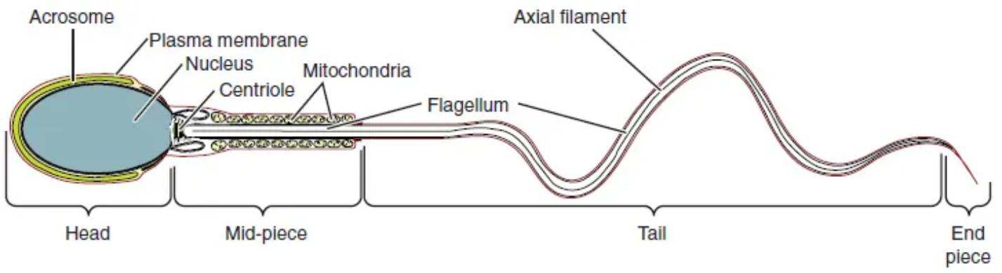
Diagram Labels and Explanations:
Acrosome: This is a cap-like organelle located at the anterior part of the sperm head. It contains hydrolytic enzymes, such as hyaluronidase and acrosin, which are essential for penetrating the outer layers of the oocyte during fertilization.
Plasma membrane: This outer lipid bilayer encloses the entire sperm cell, regulating the passage of substances and mediating interactions with the surrounding environment. It plays a critical role in capacitation and the acrosome reaction, both vital for successful fertilization.
Nucleus: Situated within the head of the sperm, the nucleus contains the highly condensed, haploid paternal DNA. Its compact nature protects the genetic material and contributes to the streamlined shape of the sperm head.
Centriole: Located at the base of the nucleus, the centriole is crucial for the formation of the flagellum during spermiogenesis. After fertilization, it also plays a vital role in the formation of the zygote’s first mitotic spindle.
Mitochondria: These organelles are found spirally arranged within the mid-piece of the sperm. They are responsible for generating ATP through oxidative phosphorylation, providing the necessary energy for the flagellum’s movement and thus for sperm motility.
Flagellum: This long, whip-like appendage extends from the mid-piece and forms the main part of the tail. Its rhythmic beating propels the sperm through the female reproductive tract towards the oocyte.
Axial filament: This is the core structure of the flagellum, also known as the axoneme, composed of microtubules arranged in a ‘9+2’ pattern. It is responsible for the bending and movement of the flagellum, enabling sperm motility.
Head: This is the anterior most part of the sperm cell, containing the nucleus and covered by the acrosome. Its primary function is to carry the paternal genetic material and facilitate penetration of the egg.
Mid-piece: Situated between the head and the tail, the mid-piece is characterized by its dense packing of mitochondria around the initial segment of the flagellum. This region serves as the power generator for sperm motility.
Tail: The longest segment of the sperm, extending from the mid-piece. It is responsible for the propulsive movement that drives the sperm forward, enabling it to travel through the female reproductive tract.
End piece: This is the very terminal portion of the sperm tail, characterized by a tapering structure. It marks the end of the axial filament and contributes to the overall length and flexibility of the flagellum.
The Functional Architecture of a Sperm Cell
The diagram of a sperm cell beautifully illustrates its highly specialized structure, which is optimized for its role in fertilization. Each region—the head, mid-piece, and tail—serves a distinct and vital purpose, ensuring the efficient delivery of paternal genetic material. The head is the most critical component for genetic contribution and initial interaction with the oocyte.
The sperm head is essentially a compact genetic package, containing the nucleus with its highly condensed haploid DNA. This condensation protects the delicate genetic material from damage during its journey. Capping the nucleus is the acrosome, an essential organelle filled with hydrolytic enzymes. These enzymes are crucial for digesting the outer layers of the egg, specifically the corona radiata and zona pellucida, allowing the sperm to penetrate and fuse with the oocyte membrane. The entire structure is enveloped by the plasma membrane, which is critical for various physiological changes, including capacitation, that occur in the female reproductive tract.
Following the head is the mid-piece, often described as the “powerhouse” of the sperm. This region is densely packed with mitochondria, spirally arranged around the initial segment of the flagellum. These mitochondria are responsible for producing the vast majority of the ATP required to fuel the vigorous whipping motion of the tail. Without this continuous energy supply, the sperm would be unable to travel the necessary distance to reach and fertilize the egg. The efficiency of these mitochondria is paramount for maintaining sperm motility and viability.
The tail constitutes the longest part of the sperm and is the primary organ of locomotion. Its core is the axial filament, a complex arrangement of microtubules (axoneme) that enables the characteristic undulating movement. The tail’s propulsive force drives the sperm through the female reproductive fluids, overcoming resistance to reach the oocyte. The rhythmic beating of the flagellum, extending through the tail and terminating in the slender end piece, is a marvel of cellular mechanics, allowing for directional movement.
Conclusion
In summary, the intricate ultrastructure of the spermatozoa, with its distinct head, mid-piece, and tail regions, is meticulously designed for its critical role in reproduction. Each component, from the enzyme-rich acrosome and DNA-bearing nucleus to the energy-generating mitochondria and the propulsive flagellum, works in concert to achieve fertilization. This highly specialized cellular architecture underscores the complexity and precision of biological design, essential for the perpetuation of species. Understanding these features is fundamental to reproductive biology and the study of male infertility.

