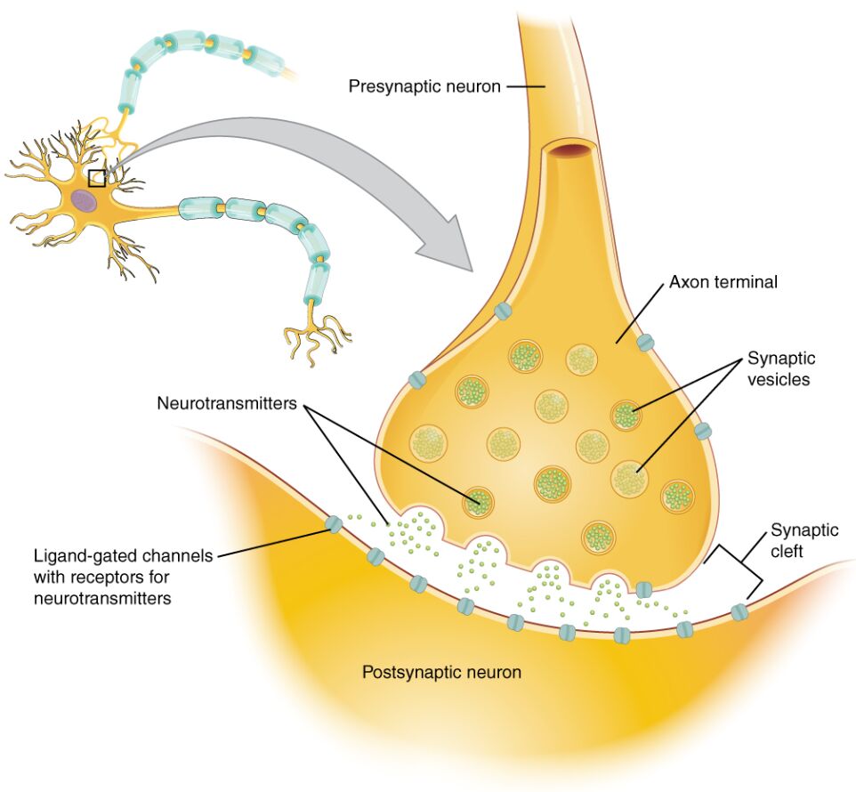
Synapses are the fundamental junctions where neurons communicate, allowing the transfer of information through chemical or electrical signals to coordinate complex bodily functions and behaviors. This diagram illustrates a chemical synapse, detailing the presynaptic and postsynaptic components involved in neurotransmitter release and reception, essential for understanding neural circuits in the brain and peripheral nervous system. By examining these elements, one gains insight into how signals propagate across the synaptic cleft, influencing everything from reflexes to memory formation.
Labeled Parts of the Synapse
Presynaptic neuron
The presynaptic neuron is the sending cell in the synaptic connection, with its axon extending to form the terminal where signals originate. It releases neurotransmitters in response to action potentials, modulating the activity of the target cell through controlled vesicle exocytosis.
Axon terminal
The axon terminal, also known as the synaptic end bulb, is the swollen end of the presynaptic axon housing synaptic vesicles and mitochondria for energy supply. Here, calcium influx triggers fusion events, ensuring precise timing in signal transmission to the postsynaptic side.
Synaptic vesicles
Synaptic vesicles are small membrane-bound sacs within the axon terminal that store neurotransmitters like glutamate or GABA, ready for release. They dock at active zones and fuse with the plasma membrane via SNARE proteins, discharging contents into the cleft upon depolarization.
Neurotransmitters
Neurotransmitters are chemical messengers depicted diffusing across the cleft, such as acetylcholine in neuromuscular junctions or serotonin in mood regulation. They bind to postsynaptic receptors to elicit excitatory or inhibitory postsynaptic potentials, with clearance mechanisms preventing prolonged signaling.
Synaptic cleft
The synaptic cleft is the narrow extracellular space, about 20-40 nm wide, separating presynaptic and postsynaptic membranes. It allows neurotransmitter diffusion while housing enzymes and transporters for rapid signal termination.
Ligand-gated channels with receptors for neurotransmitters
Ligand-gated channels with receptors for neurotransmitters are ionotropic receptors on the postsynaptic membrane that open upon binding specific molecules, allowing ion flux like Na+ for depolarization. These channels, such as AMPA for glutamate, mediate fast synaptic transmission essential for rapid neural responses.
Postsynaptic neuron
The postsynaptic neuron receives the signal, with its dendrite or soma featuring receptors that integrate inputs from multiple synapses. It processes these to determine whether to fire an action potential, contributing to network-level computations.
In-Depth Anatomy of the Chemical Synapse
The structure of a chemical synapse is highly specialized for unidirectional communication between neurons. Anatomical features ensure efficiency and specificity in signal transfer.
- The presynaptic axon terminal contains voltage-gated calcium channels (Cav2.1) clustered at active zones, facilitating rapid Ca2+ entry during depolarization.
- Synaptic vesicles, 40-50 nm in diameter, are recycled via endocytosis involving clathrin coats to maintain transmission during high-frequency firing.
- The synaptic cleft includes extracellular matrix proteins like laminins, stabilizing the junction and modulating diffusion rates.
- Postsynaptic densities, electron-dense regions, anchor receptors via scaffolding proteins such as PSD-95 for glutamate receptors.
- Overall, synapse morphology varies: axodendritic (axon to dendrite) predominate, but axosomatic or axoaxonic types allow direct modulation.
Physiological Processes in Synaptic Transmission
Synaptic function relies on electrochemical gradients and molecular interactions for precise signaling. Physiological steps from release to response enable neural plasticity.
- Action potentials depolarize the terminal, opening calcium channels and elevating intracellular Ca2+ to micromolar levels for vesicle priming.
- Neurotransmitter binding to ligand-gated channels causes conformational changes, permitting selective ion permeation (e.g., Cl- in GABA_A receptors for hyperpolarization).
- Clearance involves monoamine oxidase for degradation of catecholamines or transporters like EAAT for glutamate reuptake into glia.
- Synaptic strength modulates via long-term potentiation, involving NMDA receptor activation and AMPA insertion for memory.
- Quantal release, where each vesicle equates to a quantum, underlies miniature potentials observable in electrophysiological recordings.
Types and Variations of Synapses
Synapses exhibit diversity in form and function across the nervous system. Variations adapt to specific neural demands.
- Chemical synapses, as shown, contrast with electrical synapses via gap junctions (connexins) for bidirectional, gapless ion flow in synchronized networks like cardiac muscle.
- Excitatory synapses often use glutamate on spines, while inhibitory ones employ glycine in spinal cord for motor control.
- Neuromuscular junctions, a specialized type, feature large terminals innervating single fibers with high safety factors for reliable contraction.
- Modulatory synapses release neuropeptides like substance P alongside classical transmitters for prolonged effects.
- Developmental pruning refines synaptic density, reducing from billions in infancy to optimized adult circuits.
Molecular Components and Regulation
Molecular machinery governs synapse assembly and maintenance. Regulatory pathways ensure homeostasis and adaptation.
- Vesicle fusion relies on synaptotagmin as a calcium sensor, interacting with syntaxin and SNAP-25 in the SNARE complex.
- Receptors like metabotropic types (G-protein coupled) activate second messengers (cAMP, IP3) for slower, modulatory responses.
- Glial cells envelop synapses, with astrocytes expressing transporters (GLT-1) to buffer extracellular glutamate and prevent excitotoxicity.
- Hormonal influences, such as cortisol in stress, alter synaptic plasticity via glucocorticoid receptors on neurons.
- Epigenetic factors, including histone acetylation, regulate gene expression for synaptic proteins during learning.
Synaptic Plasticity and Learning
Synapses dynamically change to support learning and memory formation. Plasticity mechanisms underpin cognitive functions.
- Hebbian theory posits “cells that fire together wire together,” manifested in LTP with calcium-dependent kinase activation.
- LTD (long-term depression) weakens synapses via phosphatase activity, balancing network excitability.
- Homeostatic scaling adjusts receptor numbers to maintain firing rates amid varying inputs.
- Experience-dependent rewiring, as in visual cortex critical periods, involves BDNF signaling for dendritic spine growth.
- Sleep consolidates synaptic changes, with slow-wave activity downscaling overall strength for energy efficiency.
Advances in Synaptic Research
Recent methodologies enhance understanding of synaptic operations. Techniques reveal nanoscale details and dynamics.
- Super-resolution microscopy (STED) visualizes vesicle clusters and receptor trafficking in live cells.
- Optogenetics controls release with light-sensitive channels, dissecting circuit roles in behavior.
- Cryo-electron tomography reconstructs 3D synapse ultrastructure, identifying novel proteins.
- Single-cell sequencing profiles transcriptomes, uncovering synapse-type specific genes.
- Computational models simulate transmission using Hodgkin-Huxley equations extended for quantal variability.
Potential Pathologies at the Synapse
Although the diagram shows a healthy synapse, dysfunctions contribute to neurological disorders. Pathological insights drive therapeutic developments.
- Alzheimer’s involves amyloid-beta disrupting glutamate clearance, leading to excitotoxicity and synapse loss.
- Parkinson’s features dopamine synapse degeneration in substantia nigra, with levodopa restoring transmission.
- Epilepsy arises from imbalanced excitation-inhibition, often due to GABA receptor mutations.
- Schizophrenia links to altered dopamine and glutamate signaling, treated with antipsychotics targeting D2 receptors.
- Autism spectrum disorders show synaptic pruning deficits, with mTOR pathway hyperactivity affecting connectivity.
In summary, the chemical synapse depicted serves as the cornerstone of neural communication, with its labeled components orchestrating the release, reception, and clearance of neurotransmitters for seamless information flow. Continued study of these junctions not only unravels brain mysteries but also fosters innovations in treating synaptic-related conditions, enhancing neurological well-being.

