Understanding the Structures of the Respiratory Zone: Alveoli and Microscopic Views
The respiratory zone of the lungs is a critical area where gas exchange occurs, ensuring oxygen enters the bloodstream and carbon dioxide is expelled. This article delves into the intricate structures of the alveoli and their microscopic appearance, providing a comprehensive look at their anatomy and function through detailed diagrams and images.
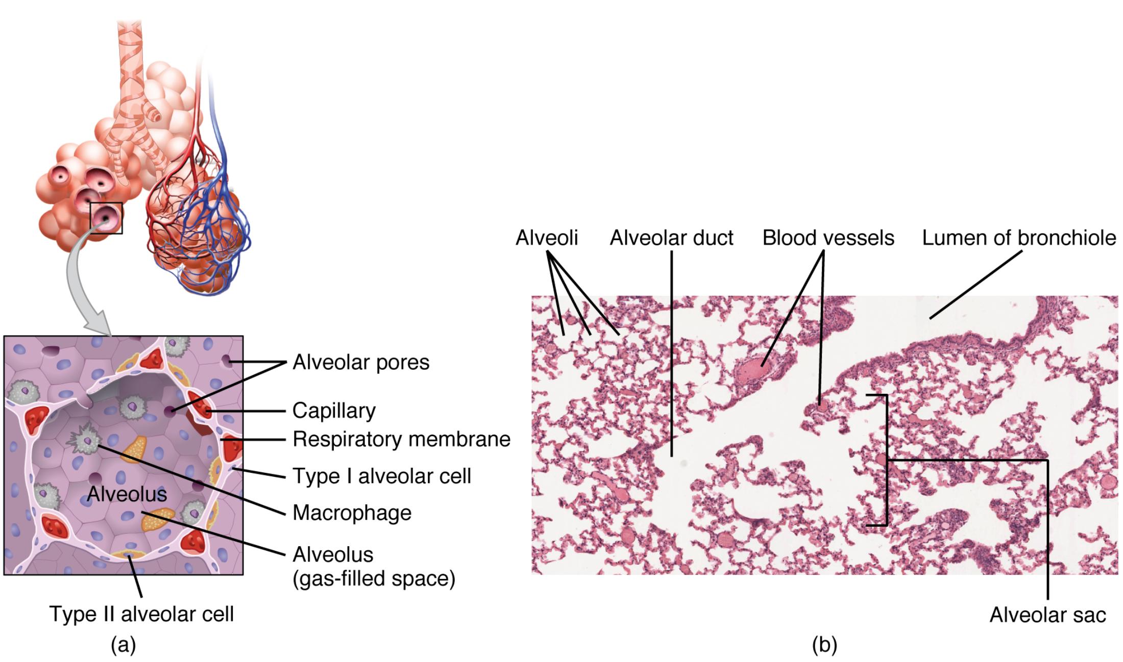
Alveoli: These tiny air sacs are the primary sites for gas exchange in the lungs, allowing oxygen to move into the blood and carbon dioxide to be removed. Their thin walls and large surface area, approximately 70-100 square meters in adults, maximize the efficiency of this process.
Alveolar duct: This small passageway connects the respiratory bronchioles to the alveoli, facilitating the flow of air to these gas exchange units. It plays a vital role in distributing air evenly across the alveolar sacs for optimal respiratory function.
Blood vessels: These capillaries surround the alveoli, enabling the close proximity needed for the diffusion of gases across the respiratory membrane. The dense network ensures a continuous supply of oxygenated blood to the body and the removal of deoxygenated blood.
Lumen of bronchiole: This central airway within the bronchiole allows air to travel from the trachea to the alveoli, serving as a conduit for inhaled and exhaled air. Its smooth muscle lining helps regulate airflow during breathing.
Alveolar pores: These small openings between adjacent alveoli allow for the equalization of air pressure and provide alternative pathways for air movement. They also assist in collateral ventilation, which can be crucial in maintaining lung function if some airways are blocked.
Capillary: These tiny blood vessels within the alveoli walls are where oxygen and carbon dioxide are exchanged, with a thickness of only about 0.5-1 micrometer to enhance diffusion efficiency. Their extensive branching increases the surface area available for gas exchange.
Respiratory membrane: This thin barrier, consisting of the alveolar wall, capillary endothelium, and their fused basement membranes, facilitates the rapid diffusion of gases. Its integrity is essential for effective oxygen uptake and carbon dioxide removal.
Type I alveolar cell: These flat, squamous cells form the majority of the alveolar lining, providing a thin surface for gas exchange due to their minimal thickness of about 0.1-0.5 micrometers. They are highly specialized to support the respiratory process without impeding diffusion.
Macrophage: These immune cells patrol the alveoli, engulfing and removing dust, pathogens, and debris to maintain a sterile environment. Their presence is crucial for preventing infections and maintaining lung health.
Alveolus (gas-filled space): This gas-filled sac within the lung tissue holds air during respiration, creating the environment for oxygen and carbon dioxide exchange. Its elastic nature allows it to expand and contract with each breath.
Type II alveolar cell: These cuboidal cells produce surfactant, a substance that reduces surface tension to prevent alveolar collapse during exhalation. They also play a role in repairing the alveolar lining if damage occurs.
Alveolar sac: This cluster of alveoli acts as a collective unit for gas exchange, increasing the total surface area for respiration. Its structure enhances the efficiency of oxygen and carbon dioxide transfer in the lungs.
Anatomical Overview of the Respiratory Zone
The respiratory zone is where the magic of breathing happens, transforming inhaled air into life-sustaining oxygen for the body. This section explores the key structures that make this process possible, starting with the alveoli and their supporting components.
- The alveoli are tiny, balloon-like structures clustered at the end of the bronchial tree, designed to maximize gas exchange.
- Surrounding these alveoli are blood vessels, particularly capillaries, which create a network for oxygen to enter the bloodstream.
- The alveolar ducts serve as conduits, channeling air from the bronchioles to the alveoli, ensuring a steady airflow.
- Within the alveoli, the gas-filled spaces are maintained by the elastic properties of the lung tissue, aiding in efficient breathing.
Microscopic Insights into Alveolar Structures
Delving deeper, the microscopic view reveals the intricate details of lung tissue, offering a closer look at cellular components. This perspective is crucial for understanding how the respiratory system functions at a cellular level.
- The micrograph shows alveoli as pink-stained structures, highlighting their abundance and interconnectedness within the lung.
- Type I alveolar cells dominate the alveolar walls, their thin structure optimized for gas diffusion across the respiratory membrane.
- Macrophages are visible as scattered cells, actively defending the alveoli against inhaled particles and pathogens.
- Type II alveolar cells, though fewer in number, are vital for producing surfactant to keep the alveoli open.
Functional Roles in Gas Exchange
The process of gas exchange is a finely tuned mechanism, relying on the structural integrity of the alveoli and their associated components. This section outlines how each part contributes to this essential function.
- The respiratory membrane’s thinness allows oxygen to diffuse into the capillaries and carbon dioxide to exit, a process occurring in mere fractions of a second.
- Alveolar pores provide a backup route for air, ensuring ventilation even if primary airways are obstructed.
- The lumen of the bronchiole regulates airflow, adjusting to the body’s oxygen demands during rest or exercise.
- Capillaries, with their extensive network, ensure that deoxygenated blood is quickly replenished with oxygen.
Conclusion
Understanding the structures of the respiratory zone, from the alveoli to the microscopic cellular level, is fundamental to appreciating how our lungs sustain life. The interplay of blood vessels, alveolar cells, and supporting ducts ensures efficient gas exchange, a process that occurs tirelessly with every breath. This detailed exploration provides a solid foundation for further study and appreciation of respiratory anatomy and physiology.
alveoli, respiratory zone, gas exchange, alveolar ducts, blood vessels, lumen of bronchiole, alveolar pores, capillaries, respiratory membrane, type I alveolar cells, macrophages, type II alveolar cells, alveolar sac, lung tissue, surfactant

