Prokaryotic ribosomes are the essential protein-manufacturing machines found within bacterial cells. Unlike eukaryotic cells, bacteria utilize a 70S ribosome composed of two distinct subunits, which serve as a critical target for many lifesaving antibiotics. Understanding the precise anatomical structure of these ribosomal components is fundamental to both molecular biology and clinical pharmacology.
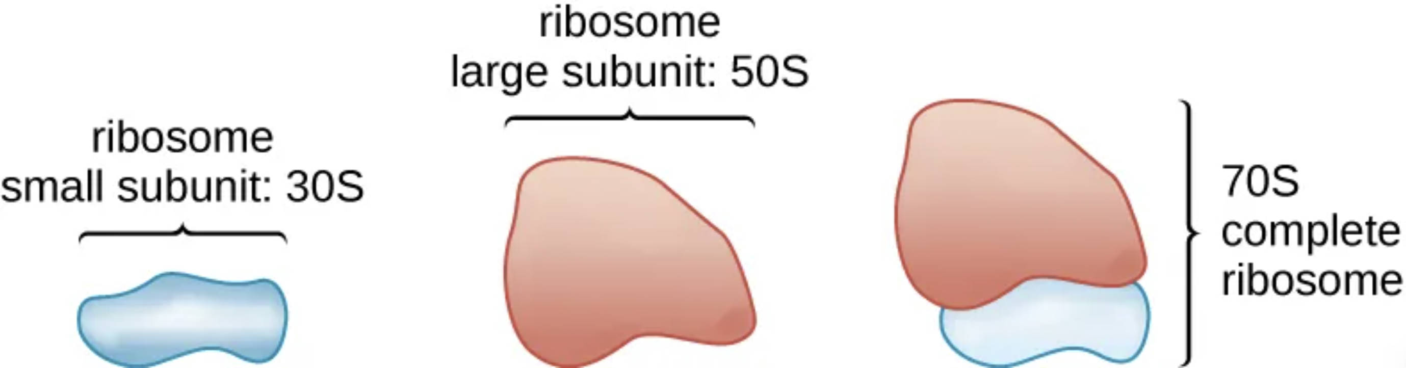
ribosome small subunit: 30S: This smaller portion of the ribosome is responsible for reading the genetic code carried by messenger RNA (mRNA) during the initiation of protein synthesis. It is composed of a single 16S ribosomal RNA (rRNA) molecule and approximately 21 different proteins that stabilize the structure.
ribosome large subunit: 50S: The larger component of the prokaryotic ribosome catalyzes the formation of peptide bonds between amino acids to build a protein chain. It contains the 5S and 23S rRNA molecules along with roughly 34 proteins, forming the complex where the actual biochemical assembly occurs.
70S complete ribosome: This is the functional unit formed when the 30S and 50S subunits join together onto an mRNA strand to begin building a polypeptide. Its name refers to its sedimentation rate in Svedberg units, a measurement that depends on both the mass and the three-dimensional surface area of the assembled machine.
The Biological Workhorses: Ribosomes in Prokaryotic Life
Ribosomes are the ubiquitous “workhorses” of the cell, responsible for the vital process of converting genetic blueprints into the proteins that drive life. In prokaryotes—organisms like bacteria that lack a membrane-bound nucleus—these ribosomes are scattered throughout the cytoplasm, ready to engage in protein synthesis as soon as messenger RNA is transcribed. This immediate accessibility is one of the reasons bacteria can replicate so quickly, making them formidable survivors in diverse environments.
The classification of ribosomes as “70S” is a common point of confusion for students, as the math of 30S and 50S doesn’t seem to add up. However, sedimentation rates are not additive because they are influenced by the shape and density of the particle as it moves through a liquid medium during centrifugation. When the two subunits dock together, they create a more compact and streamlined functional unit that settles at the 70S mark.
The structural complexity of these machines is what allows them to manage the intricate choreography of translation. Essential functions include:
- Recognition of start codons on mRNA strands to ensure correct protein coding.
- Provision of structural scaffolding for transfer RNA (tRNA) molecules carrying amino acids.
- Catalysis of peptide bond formation through the ribozyme activity of the 23S rRNA.
- Translocation along the mRNA template to read the next genetic instruction.
In the medical field, the subtle structural differences between the 70S bacterial ribosome and the 80S human ribosome are of paramount importance. This distinction is the foundation of selective toxicity, allowing clinicians to prescribe medications that kill invading bacteria without harming the patient’s own healthy cells.
Clinical Pharmacology and Ribosomal Targeting
Because bacteria cannot survive without the ability to produce proteins, the 70S ribosome is one of the most effective targets in antimicrobial therapy. Many classes of antibiotics are designed to bind to specific sites on either the 30S or 50S subunit. For example, aminoglycosides such as gentamicin bind to the 30S subunit, causing the ribosome to misread the mRNA, leading to the production of faulty proteins that ultimately kill the bacterium.
Other drugs, like macrolides (e.g., erythromycin), target the 50S subunit. These molecules physically block the “exit tunnel” where the growing protein chain emerges, effectively stalling the ribosome and preventing the bacteria from growing. Understanding these microscopic interactions is vital for managing drug resistance, as bacteria often try to mutate the shape of their ribosomal subunits or the associated rRNA to prevent antibiotics from binding.
The elegant design of the prokaryotic ribosome highlights the efficiency of life at its most basic level. By coordinating the interaction of RNA and proteins, these 70S machines ensure that the genetic instructions of the cell are accurately realized as functional structures. Continued research into the structural biology of the ribosome remains a cornerstone of modern medicine, providing the necessary insights to develop the next generation of life-saving antibacterial treatments.

