Explore the remarkable histological adaptations of the small intestine that dramatically enhance its capacity for nutrient absorption. This article delves into the intricate structure of circular folds, villi, and microvilli, detailing how these features collectively create an enormous surface area essential for efficient digestion and nutrient uptake. Understanding this microscopic architecture is fundamental to comprehending the small intestine’s critical role in human health.
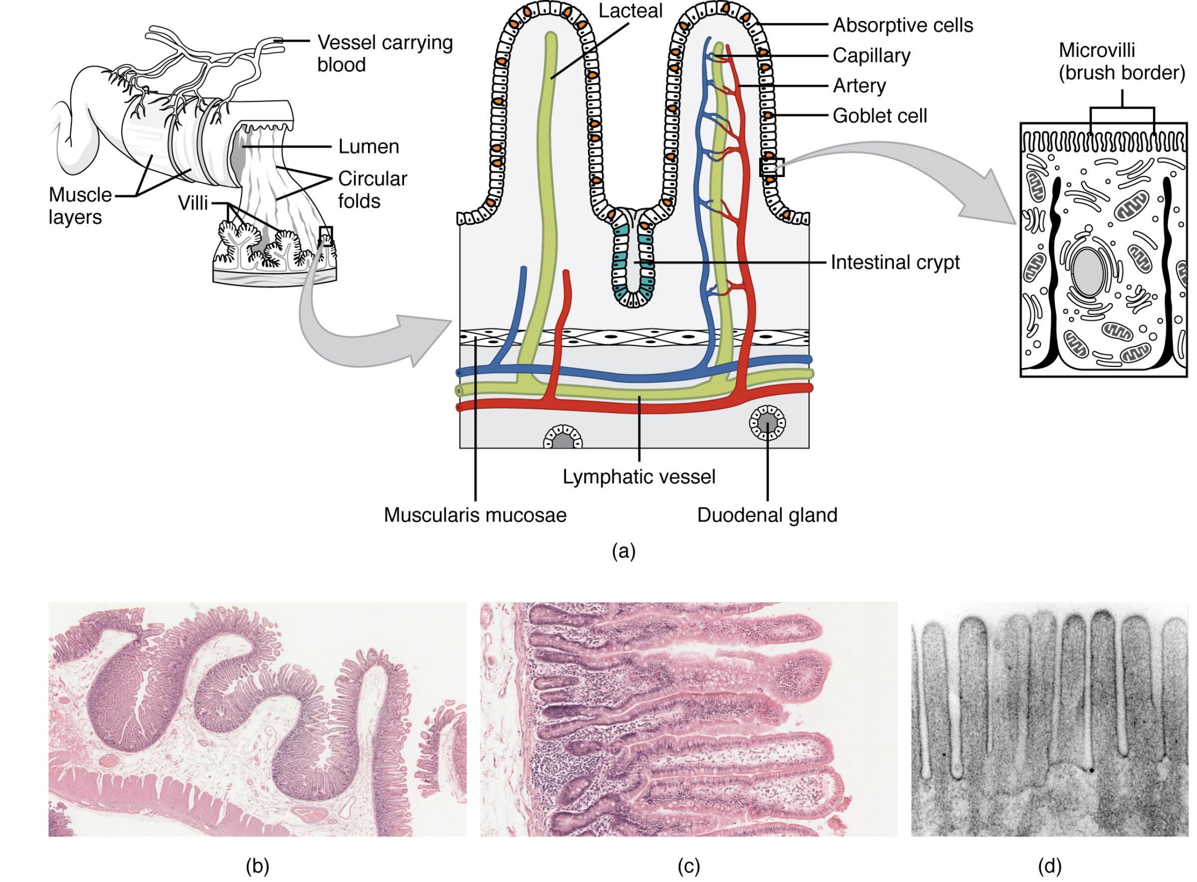
Vessel carrying blood: These are the arteries and veins that supply oxygenated blood and remove deoxygenated blood and absorbed nutrients from the small intestine. They form a dense network within the villi, facilitating the rapid transport of absorbed substances.
Muscle layers: These layers of smooth muscle are responsible for the movements of the small intestine, including segmentation and peristalsis. These contractions help mix chyme with digestive juices and propel it along the digestive tract.
Lumen: This refers to the inner open space within the small intestine through which partially digested food, or chyme, passes. It is where digestion occurs and absorbed nutrients interact with the intestinal lining.
Circular folds: Also known as plicae circulares, these are large, crescent-shaped folds of the small intestine’s mucosal and submucosal layers. They significantly increase the surface area and slow down the passage of chyme, allowing more time for digestion and absorption.
Villi: These are finger-like projections of the intestinal mucosa that extend into the lumen, giving the inner surface a velvety appearance. Each villus is covered with absorptive cells and contains a rich capillary network and a lacteal for nutrient transport.
Lacteal: A lymphatic capillary located within the core of each villus. Lacteals are specifically responsible for absorbing digested fats (chylomicrons), which are then transported into the lymphatic system.
Absorptive cells: Also known as enterocytes, these are the primary cells lining the villi, specialized for the absorption of nutrients. They possess numerous microvilli on their apical surface, forming the brush border.
Capillary: A tiny blood vessel forming a network within each villus, intimately associated with the absorptive cells. These capillaries absorb most digested nutrients, such as amino acids and monosaccharides, directly into the bloodstream.
Artery: A blood vessel that carries oxygenated blood and nutrients to the tissues of the small intestine, including the villi and crypts. It branches into smaller arterioles and then capillaries to facilitate exchange.
Goblet cell: These specialized glandular cells are scattered among the absorptive cells within the intestinal lining. Their primary function is to secrete mucus, which lubricates and protects the intestinal surface from enzymatic digestion and mechanical abrasion.
Intestinal crypt: Also known as the crypts of Lieberkühn, these are invaginations of the intestinal epithelium located between the bases of the villi. They contain various cell types, including stem cells that continuously regenerate the intestinal lining, as well as enteroendocrine cells and Paneth cells.
Lymphatic vessel: Part of the lymphatic system, these vessels run through the submucosa and receive lymph from the lacteals. They play a crucial role in immune surveillance and the transport of absorbed fats.
Muscularis mucosae: A thin layer of smooth muscle separating the mucosa from the submucosa. Its contractions help to move the villi, enhancing contact with digested food and facilitating absorption.
Duodenal gland: Also known as Brunner’s glands, these are compound tubular glands found specifically in the submucosa of the duodenum. They secrete an alkaline mucus rich in bicarbonate, which helps neutralize the acidic chyme entering from the stomach, protecting the duodenal lining.
Microvilli (brush border): These are microscopic, hair-like projections extending from the apical surface of each absorptive cell. They form a dense “brush border” that vastly increases the surface area for enzyme activity and nutrient absorption, representing the final stage of surface amplification.
The small intestine is a remarkable organ, meticulously engineered to perform the crucial task of nutrient absorption. While its considerable length (approximately 6-7 meters in adults) contributes to its capacity, it is the extraordinary folding of its internal surface that truly maximizes its digestive and absorptive efficiency. This intricate architectural design ensures that virtually every available nutrient from digested food is captured and transported into the bloodstream or lymphatic system.
The hierarchical organization of these folds provides a staggering increase in surface area. Imagine a smooth tube; the small intestine, however, is anything but smooth. It employs three distinct levels of folding:
- Circular folds (plicae circulares), which are macroscopic ridges of the mucosa and submucosa.
- Villi, which are finger-like projections extending from these folds.
- Microvilli, microscopic projections on the surface of individual absorptive cells, forming what is known as the brush border.
These structural adaptations are not merely for show; they are vital for the efficient breakdown and absorption of carbohydrates, proteins, and fats. Without this vast surface area, the process of nutrient uptake would be significantly compromised, leading to malabsorption and nutritional deficiencies. Each level of folding plays a specific role, working in concert to create an optimal environment for digestion.
The histology of the small intestine reveals a landscape optimized for absorption, a testament to the sophistication of biological design. The inner lining, or mucosa, is not merely a simple barrier but a dynamic, highly structured interface where most nutrient assimilation occurs.
Circular Folds: Macroscopic Amplifiers
The circular folds, visible to the naked eye, are permanent, transverse ridges of the mucosa and submucosa. These folds begin in the proximal duodenum and are most numerous in the jejunum, gradually decreasing in size and number towards the ileum. Their primary function is to increase the surface area by a factor of approximately three, and crucially, they also cause the chyme to spiral through the intestine, slowing its passage and ensuring maximum contact with the absorptive surface.
Villi: Finger-like Projections for Absorption
Superimposed on the circular folds are the villi, which are small, finger-like or leaf-like projections of the mucosal layer that extend into the lumen. Each villus is approximately 0.5-1.5 mm long and is covered by a layer of epithelial cells, primarily absorptive enterocytes and mucus-secreting goblet cells. Within the core of each villus lies a rich vascular supply, including a dense capillary network for absorbing water-soluble nutrients (monosaccharides, amino acids, vitamins, minerals) directly into the bloodstream. A central lymphatic capillary, known as a lacteal, is specifically responsible for absorbing digested fats, which are too large to enter the capillaries directly. The villi increase the surface area by roughly tenfold.
Microvilli: The Brush Border’s Microscopic Detail
The most significant amplification of the absorptive surface comes from the microvilli, which are minute, hair-like projections on the apical (lumen-facing) surface of each absorptive cell (enterocyte). These microscopic structures form a dense, fuzzy layer known as the brush border. Enzymes embedded within the brush border complete the final stages of carbohydrate and protein digestion, breaking them down into their simplest forms ready for absorption. The microvilli increase the surface area by an astonishing twentyfold, bringing the total effective absorptive surface of the small intestine to an area comparable to a tennis court. This intricate design ensures maximum efficiency in nutrient uptake.
In summary, the small intestine’s ability to efficiently absorb nutrients is a direct consequence of its remarkable histological architecture. The sequential layering of circular folds, villi, and microvilli dramatically expands the available surface area, allowing for extensive contact between the chyme and the absorptive cells. This sophisticated design, supported by a rich network of blood and lymphatic vessels within each villus, is a cornerstone of our digestive health. Understanding these microscopic features provides profound insight into how our bodies acquire the vital nutrients necessary for life.

