Explore the vital connection between erythrocytes (red blood cells) and hemoglobin, the protein responsible for oxygen transport throughout the body, as illustrated in this detailed diagram. This article delves into the intricate structure of hemoglobin, highlighting its four subunits and their crucial iron-containing core. Gain a deeper understanding of how these microscopic components are essential for cellular respiration and overall physiological health.
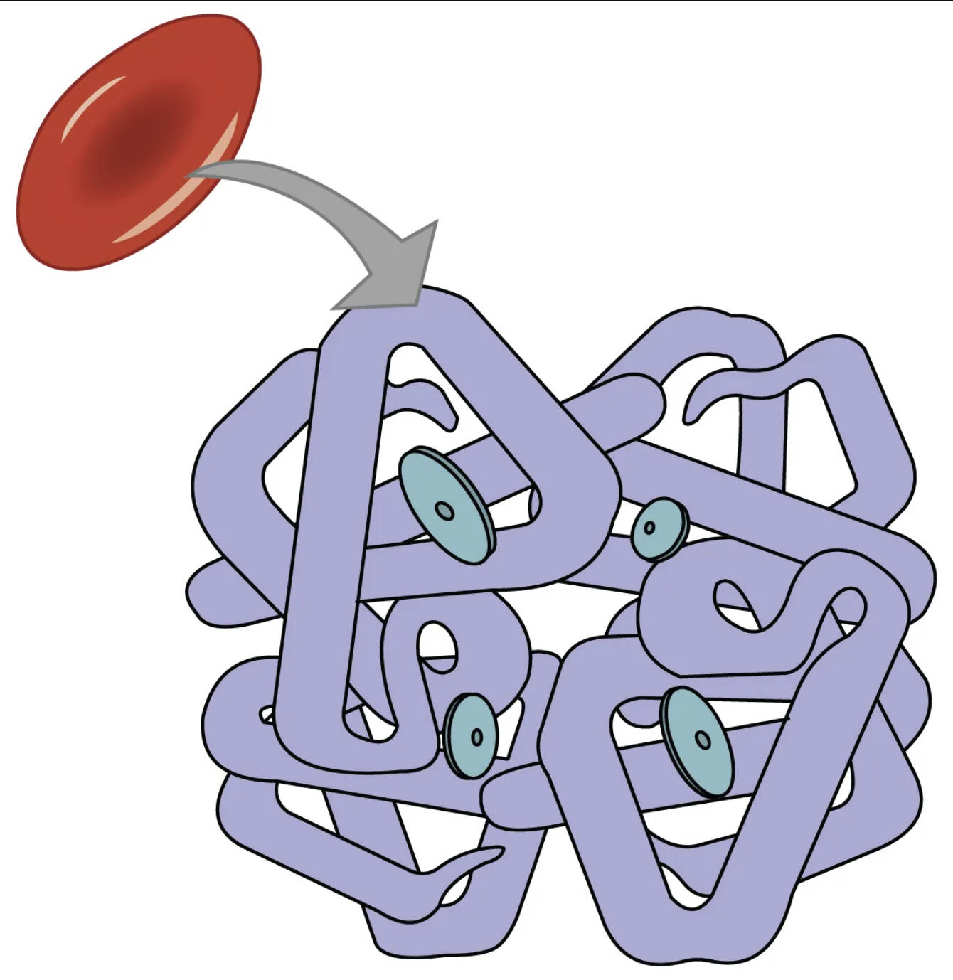
The efficient transport of oxygen from the lungs to every cell in the body is a fundamental physiological process, primarily orchestrated by red blood cells, or erythrocytes. Within these biconcave disc-shaped cells resides a specialized protein called hemoglobin, which is exquisitely designed for binding and releasing oxygen. Hemoglobin’s unique structure allows it to pick up oxygen where it is abundant (in the lungs) and deliver it to tissues where oxygen levels are lower (in metabolically active cells). A comprehensive understanding of this molecule is central to comprehending respiratory physiology and various hematological conditions. The diagram vividly illustrates an erythrocyte and the complex, globular structure of a hemoglobin molecule, emphasizing its multi-subunit composition.
The Structure of Hemoglobin
Hemoglobin is a quaternary protein, meaning it is composed of multiple polypeptide chains. Specifically, it consists of four protein subunits: two alpha (α) globin chains and two beta (β) globin chains in adult hemoglobin (HbA). Each of these four globin subunits is non-covalently bound to a heme group.
- Four Subunits: As indicated, hemoglobin is made up of four protein subunits. In adult hemoglobin (HbA), these are typically two alpha-globin chains and two beta-globin chains. The precise arrangement of these subunits creates a stable, functional protein.
- Each of which contains one molecule of iron: This refers to the critical iron atom located at the center of each heme group within each globin subunit. It is this ferrous iron (Fe2+) that reversibly binds to oxygen, allowing hemoglobin to transport oxygen. Each hemoglobin molecule can, therefore, carry up to four molecules of oxygen.
The Role of Erythrocytes in Oxygen Transport
Erythrocytes are specialized cells, lacking a nucleus and most organelles in their mature form, which maximizes their capacity for hemoglobin. Their biconcave shape increases their surface area-to-volume ratio, facilitating efficient gas exchange. As red blood cells circulate, hemoglobin within them binds to oxygen in the pulmonary capillaries, forming oxyhemoglobin. This oxygenated blood is then pumped by the heart to systemic capillaries, where hemoglobin releases oxygen to meet the metabolic demands of tissues, becoming deoxyhemoglobin. Concurrently, hemoglobin also plays a role in transporting a small amount of carbon dioxide and buffering pH in the blood.
Clinical Significance of Hemoglobin
Disruptions in hemoglobin production or structure can lead to various medical conditions. For instance, anemia often results from insufficient hemoglobin or red blood cells, leading to reduced oxygen-carrying capacity and symptoms like fatigue and shortness of breath. Genetic disorders such as sickle cell anemia or thalassemia involve abnormal globin chains, which impair hemoglobin function and erythrocyte integrity. Iron deficiency, a common nutritional deficit, directly impacts hemoglobin synthesis, as iron is a crucial component of the heme group. Monitoring hemoglobin levels is a standard diagnostic tool in medicine, providing insights into a patient’s oxygen transport capabilities.
In conclusion, the intricate relationship between erythrocytes and the hemoglobin protein is fundamental to life. Hemoglobin’s quaternary structure, comprising four globin subunits, each with a central iron molecule, enables the efficient and reversible transport of oxygen throughout the body. Understanding this vital molecular and cellular mechanism is essential not only for appreciating normal physiological function but also for diagnosing and managing a wide array of hematological disorders that affect oxygen delivery and overall health.

