The olfactory system, a cornerstone of human sensory perception, originates in the nasal cavity where it captures and processes odor molecules from the environment. This image illustrates the key components, including the olfactory epithelium and its receptor neurons, highlighting the initial stages of smell detection and neural transmission. This article provides a detailed exploration of the anatomy and physiology of these structures, offering valuable insights into their roles in the olfactory process.
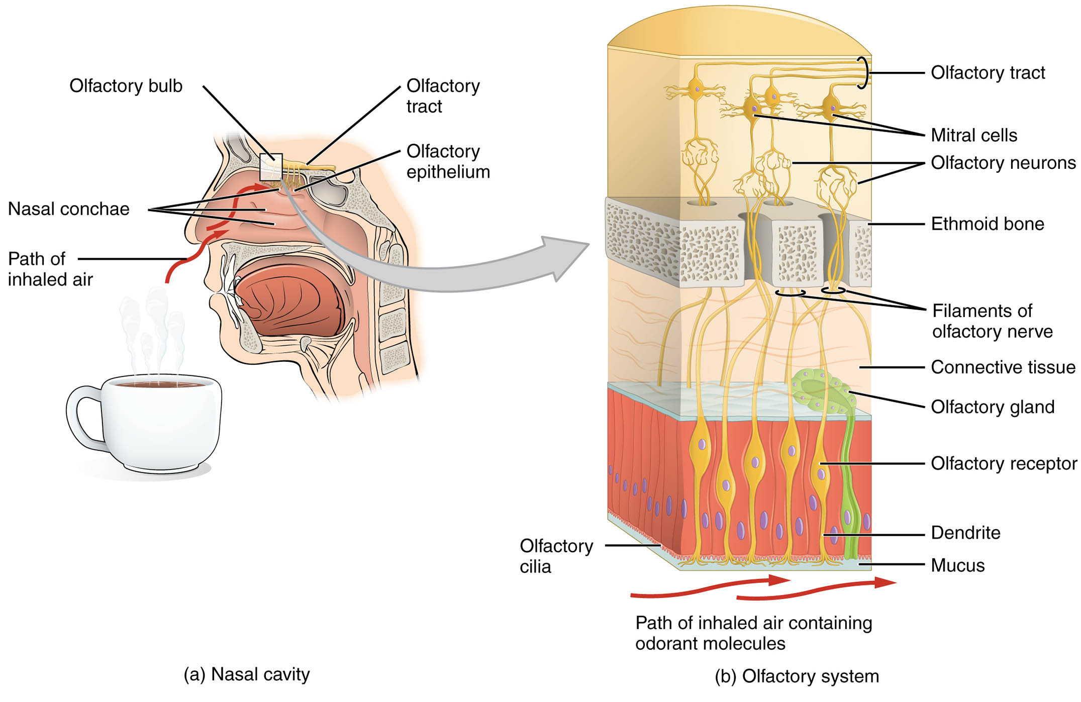
Labeled Parts of the Olfactory System
Olfactory epithelium The olfactory epithelium is a specialized tissue lining the upper nasal cavity, containing olfactory receptor neurons that detect odorants. It is supported by a mucus layer and connective tissue, creating an optimal environment for sensory function.
Olfactory receptor neuron The olfactory receptor neuron is a bipolar cell within the olfactory epithelium, with dendrites equipped with cilia to detect odors and an axon to transmit signals. Its role is critical in converting chemical stimuli into neural impulses for the brain.
Cilia Cilia are hair-like extensions on the dendrites of olfactory receptor neurons, increasing the surface area for odorant molecule binding. They move within the mucus, enhancing the efficiency of smell detection by facilitating odorant-receptor interactions.
Mucus layer The mucus layer covers the olfactory epithelium, dissolving odorant molecules to enable their detection by receptor neurons. It is secreted by underlying glands, protecting the epithelium while supporting the olfactory process.
Supporting cell The supporting cell, or sustentacular cell, provides structural and metabolic support to olfactory receptor neurons within the epithelium. It also contributes to the mucus layer and detoxifies harmful substances, safeguarding sensory function.
Basal cell The basal cell is a stem cell at the base of the olfactory epithelium, responsible for regenerating olfactory receptor neurons. Its continuous division ensures the epithelium remains functional despite regular cell turnover.
Axon The axon is the long projection of an olfactory receptor neuron that carries electrical impulses to the olfactory bulb. These axons bundle into the olfactory nerve, forming the pathway for smell signals to reach the brain.
Nasal cavity The nasal cavity is the air-filled space where the olfactory system begins, housing the olfactory epithelium and guiding inhaled air. Its structure, including turbinates, enhances air exposure to the epithelium for odor detection.
Nasal turbinates Nasal turbinates are bony structures within the nasal cavity that increase air turbulence and surface area. They direct airflow toward the olfactory epithelium, optimizing the delivery of odorant molecules.
Anatomical Overview of the Olfactory System
The olfactory system’s structure within the nasal cavity forms the foundation for smell perception, with each component playing a specific role. This anatomical arrangement ensures efficient detection and transmission of olfactory signals.
- Epithelial location: The olfactory epithelium lines the upper nasal cavity, strategically positioned to intercept inhaled air containing odorants.
- Receptor distribution: Olfactory receptor neurons are densely packed within the epithelium, with cilia extending into the mucus layer for maximum sensitivity.
- Supportive framework: Supporting cells and basal cells maintain the epithelium’s integrity, while the mucus layer protects and activates the sensory process.
- Neural pathway: Axons from receptor neurons extend toward the olfactory bulb, with the nasal cavity serving as the entry point for this journey.
- Airflow enhancement: Nasal turbinates increase air contact with the epithelium, ensuring effective odorant delivery and detection.
Physiological Functions of the Olfactory System
The olfactory system transforms chemical cues in the nasal cavity into sensory experiences, playing a vital role in daily life. Its physiological mechanisms are designed to handle diverse odorant molecules efficiently.
- Odorant capture: Cilia on olfactory receptor neurons bind to odorant molecules dissolved in the mucus, initiating the sensory response.
- Signal generation: This binding triggers a G-protein-coupled pathway, producing electrical impulses that travel along the neuron’s axon.
- Supportive maintenance: Supporting cells nourish receptor neurons and maintain the mucus layer, ensuring optimal sensory conditions.
- Regeneration process: Basal cells replace damaged neurons, sustaining the epithelium’s sensory capability over time.
- Signal transmission: Axons carry impulses to the olfactory bulb, where initial processing occurs before further brain integration.
Developmental and Cellular Dynamics
The olfactory system develops early in fetal life, with its structures maturing to support smell perception by birth. Cellular turnover and adaptation ensure its functionality throughout life.
- Embryonic origins: The olfactory epithelium forms from ectodermal tissue, with receptor neurons and supporting cells differentiating during gestation.
- Cellular renewal: Basal cells generate new neurons and supporting cells, driven by local signaling within the epithelium.
- Ciliary development: Cilia form on neuron dendrites, enhancing detection as the system matures postnatally.
- Nasal cavity growth: The nasal cavity and turbinates develop to optimize airflow, reflecting an evolutionary adaptation for olfaction.
- Age-related changes: Sensory decline may occur with age, potentially due to reduced epithelial cell efficiency or neuronal loss.
Clinical Relevance and Olfactory Considerations
Understanding the olfactory system’s structure is essential for diagnosing and managing smell-related conditions. Clinical assessments often target these components to identify dysfunction.
- Anosmia: Loss of smell can result from damage to olfactory receptor neurons or the epithelium, often due to head trauma or viral infections.
- Hyposmia: Reduced smell sensitivity may indicate aging or neurodegenerative diseases, affecting quality of life and safety.
- Diagnostic methods: Olfactory testing with odor identification and nasal cavity imaging assess system health.
- Therapeutic approaches: Treatments include addressing inflammation, using olfactory training, or supporting regeneration with nutrients.
- Associated impacts: Impaired olfaction can affect taste perception and hazard detection, emphasizing the need for early intervention.
In conclusion, the olfactory system’s structure within the nasal cavity represents a remarkable integration of anatomy and physiology, enabling the detection and interpretation of odors. From the receptor-rich olfactory epithelium to the airflow-enhancing nasal turbinates, this system underscores the body’s ability to adapt to its sensory environment. Exploring these components provides valuable insights into sensory health and potential clinical applications.

