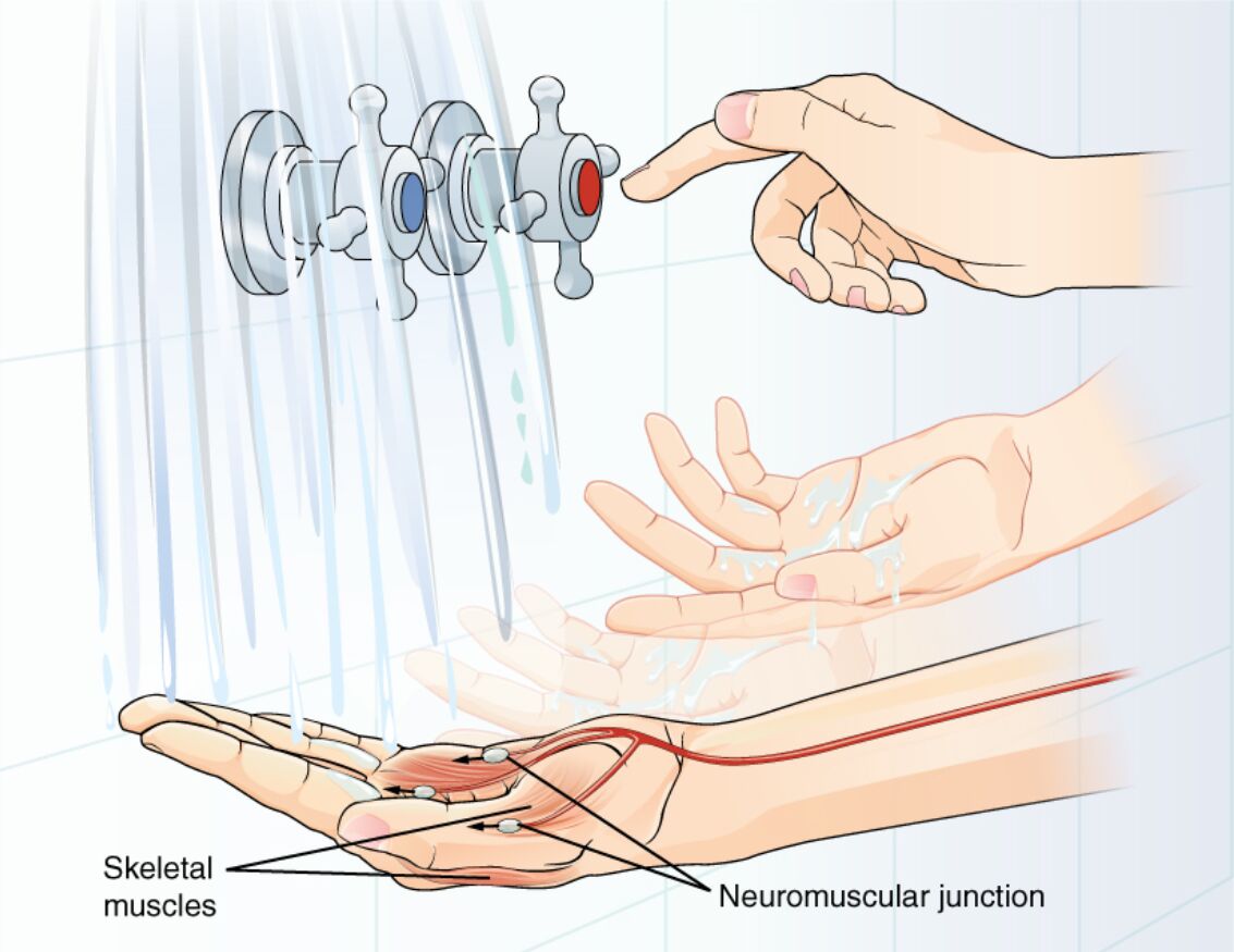
The motor response exemplifies the nervous system’s ability to translate sensory information into coordinated actions, such as withdrawing a hand from hot water after initial contact with a faucet. This diagram illustrates the sequence from stimulus detection to muscle activation, focusing on the neuromuscular junction where neural signals interface with skeletal muscles to produce movement. Such processes underscore the integration within the central nervous system (CNS), enabling both reflex and voluntary responses that protect the body and facilitate interaction with the environment.
Labeled Components in the Motor Response
Skeletal muscles
The skeletal muscles are depicted in the lower part of the hand and forearm, consisting of striated fibers responsible for voluntary movements like flexion or extension. These muscles contract in response to neural input, generating force through actin-myosin interactions within sarcomeres to execute the withdrawal action.
Neuromuscular junction
The neuromuscular junction is labeled at the interface between the motor neuron axon and muscle fiber, serving as the synapse where acetylcholine is released to trigger depolarization. This specialized structure ensures precise signal transmission, with postjunctional folds amplifying receptor density for efficient excitation-contraction coupling.
In-Depth Anatomy of the Motor Response System
The anatomy involved spans from peripheral nerves to muscle tissues, optimized for rapid and controlled actions. Key structures facilitate seamless neural-muscular communication.
- Lower motor neurons originate in the spinal cord ventral horn, their axons forming motor units that innervate groups of muscle fibers for graded force output.
- The neuromuscular junction features presynaptic terminals with voltage-gated calcium channels, synaptic clefts of 50 nm, and postsynaptic nicotinic receptors clustered by rapsyn.
- Skeletal muscles comprise fascicles of multinucleated fibers, with T-tubules and sarcoplasmic reticulum for calcium release during contraction.
- Sensory feedback from muscle spindles and Golgi tendon organs loops back via Ia/II afferents, modulating response via spinal interneurons.
- Blood supply via capillaries ensures oxygen delivery, with type I slow-twitch fibers in postural muscles contrasting type II fast-twitch in phasic ones.
Physiological Mechanisms of Muscle Contraction
Physiological events at the junction convert electrical signals to mechanical work through biochemical cascades. These mechanisms ensure timely and efficient responses.
- Action potentials arriving at axon terminals trigger calcium influx, promoting SNARE-mediated vesicle fusion and acetylcholine diffusion across the cleft.
- Binding to receptors generates end-plate potentials, spreading via sodium channels to initiate muscle action potentials along the sarcolemma.
- Calcium release from sarcoplasmic reticulum binds troponin, exposing myosin-binding sites on actin for cross-bridge cycling and shortening.
- ATP hydrolysis powers the power stroke, with relaxation occurring via SERCA pumps sequestering calcium back into storage.
- Motor unit recruitment follows Henneman’s size principle, starting with small units for fine control before larger for strength.
Integration in the Central Nervous System
CNS integration synthesizes sensory data to formulate motor commands, bridging perception and action. This process involves hierarchical neural circuits.
- Upper motor neurons in the precentral gyrus project via corticospinal tracts, decussating at the medulla to control contralateral muscles.
- Spinal cord circuits include Renshaw cells for recurrent inhibition, preventing overexcitation during responses.
- Basal ganglia modulate initiation, with dopamine facilitating smooth execution via direct pathways.
- Cerebellar input refines timing, using Purkinje cells to adjust for errors in ongoing movements.
- Cortical planning incorporates somatosensory cortex feedback for adaptive adjustments, like altering grip on slippery surfaces.
Reflex vs. Voluntary Motor Responses
Responses range from automatic reflexes to deliberate actions, differing in circuitry and speed. Distinctions highlight neural flexibility.
- Withdrawal reflexes, polysynaptic, involve spinal interneurons bypassing higher centers for sub-second latencies.
- Voluntary movements engage prefrontal areas for intention, with premotor cortex sequencing complex actions like turning a faucet.
- Both utilize alpha motor neurons as the final pathway, but reflexes incorporate flexor activation with extensor inhibition via reciprocal innervation.
- Habituation reduces reflex amplitude over time, while learning strengthens voluntary pathways through long-term potentiation.
- Pathophysiological shifts, like spasticity, arise from lost upper neuron inhibition, exaggerating reflexes.
Developmental and Molecular Aspects
Development shapes motor systems from embryonic stages, with molecular cues guiding connectivity. These foundations support lifelong function.
- Neural tube ventralization by sonic hedgehog induces motor neuron progenitors, migrating to form pools innervating specific muscles.
- Agrin from neuron terminals clusters acetylcholine receptors at junctions, stabilized by MuSK signaling.
- Postnatal myelination by Schwann cells accelerates conduction, critical for refined movements in growing limbs.
- Thyroid hormones like T3 and T4 promote neuromuscular maturation, influencing fiber type differentiation.
- Genetic expression of myogenic factors like MyoD drives muscle development, ensuring alignment with neural innervation.
Research Techniques in Motor Physiology
Investigative methods elucidate response dynamics at cellular and systemic levels. Techniques provide insights into normal and dysfunctional states.
- Electromyography (EMG) records muscle electrical activity, quantifying recruitment patterns during tasks.
- Transcranial magnetic stimulation (TMS) excites cortical neurons, measuring motor evoked potentials for pathway integrity.
- Confocal microscopy visualizes junction ultrastructure, labeling vesicles with FM dyes.
- Optogenetics in models selectively activates motor neurons, observing behavioral outputs.
- Biomechanical modeling simulates force generation, incorporating Hill’s muscle model for predictions.
Clinical Significance and Related Pathologies
Though the image portrays typical function, impairments affect daily activities, informing clinical approaches. Disorders often target junctions or pathways.
- Myasthenia gravis autoantibodies block receptors, causing fatigue treatable with acetylcholinesterase inhibitors like pyridostigmine.
- Botulism toxin inhibits acetylcholine release, leading to paralysis, countered by antitoxins.
- Muscular dystrophies like Duchenne disrupt dystrophin, weakening contraction and progressing to wheelchair dependence.
- Spinal muscular atrophy degenerates lower neurons via SMN1 mutations, with nusinersen enhancing survival.
- Traumatic injuries sever axons, but rehabilitation promotes sprouting for partial recovery.
In conclusion, the depicted motor response integrates sensory cues with neural commands to activate skeletal muscles via the neuromuscular junction, illustrating the body’s responsive elegance. Delving into these elements not only clarifies physiological harmony but also aids in developing therapies for motor disorders, advancing neurological health.

