Exploring the Lymph Node: Anatomy and Function in the Lymphatic System
The lymph node serves as a vital checkpoint in the lymphatic system, filtering lymph fluid and orchestrating immune responses to protect the body from infections and diseases. This detailed diagram offers a close-up view of its structure, highlighting the intricate network of vessels and cells that work together to maintain health and immunity.
Key Labeled Components in the Lymph Node Diagram
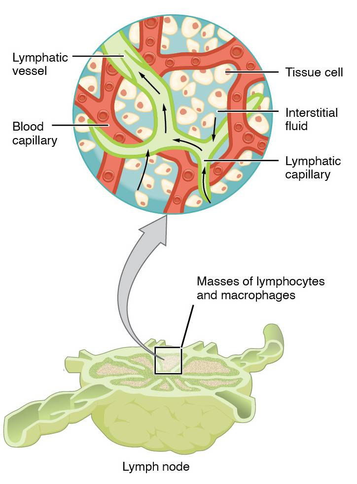
Lymphatic vessel The lymphatic vessel transports lymph fluid into and out of the lymph node, ensuring a continuous flow for filtration and immune cell interaction. These vessels are equipped with valves to prevent backflow, directing lymph toward larger ducts like the thoracic duct.
Blood capillary The blood capillary supplies oxygen and nutrients to the lymph node while allowing immune cells to enter from the bloodstream. This network facilitates the exchange of substances, supporting the node’s role in immune surveillance.
Tissue cell Tissue cells within the lymph node contribute to its structural integrity and interact with immune cells to detect pathogens. These cells are bathed in interstitial fluid, aiding in the local immune response.
Interstitial fluid Interstitial fluid fills the spaces between cells, carrying nutrients and waste while serving as the medium where pathogens are encountered by immune cells. It is collected by lymphatic capillaries to become lymph, initiating the filtration process.
Lymphatic capillary The lymphatic capillary is a thin, permeable vessel that absorbs interstitial fluid and immune cells from surrounding tissues. Its open-ended structure allows easy entry of pathogens, marking the starting point of lymph drainage.
Masses of lymphocytes and macrophages Masses of lymphocytes and macrophages form the core of the lymph node’s immune activity, with lymphocytes producing antibodies and macrophages engulfing debris. These clusters expand during infections, enhancing the node’s defensive capabilities.
Lymph node The lymph node, depicted in sectional view, acts as a filtration station where lymph is cleansed of pathogens and abnormal cells. Its internal structure includes distinct regions like the cortex and medulla, housing various immune cells for coordinated responses.
Role of the Lymph Node in Immune Defense
The lymph node plays a pivotal role in bolstering the body’s defenses. Its strategic placement along lymphatic vessels ensures constant monitoring of bodily fluids.
- Filtration Process: Lymph entering the node passes through sinuses where macrophages trap bacteria, viruses, and cellular debris, preventing systemic spread.
- Immune Cell Activation: Lymphocytes, including B-cells and T-cells, proliferate here upon detecting antigens, initiating both humoral and cell-mediated immunity.
- Antibody Production: B-cells mature into plasma cells within the node, secreting antibodies to neutralize specific pathogens.
This process is dynamic, with lymph nodes swelling during infections as immune activity intensifies. The node’s architecture, with its high density of immune cells, supports rapid responses to threats.
Microscopic Structure and Cellular Interactions
The lymph node’s internal layout reveals a complex interplay of cells and fluids. This organization enhances its efficiency in immune processing.
- Cortex and Medulla: The outer cortex contains densely packed lymphocytes in follicles, while the inner medulla features medullary cords with macrophages and plasma cells, optimizing filtration and antibody release.
- Sinuses and Reticular Network: Lymph flows through subcapsular and trabecular sinuses, supported by a reticular framework that traps pathogens for immune cell access.
- Endothelial Support: High endothelial venules within the node allow circulating lymphocytes to enter, reinforcing local immune responses.
The interaction between blood capillaries and lymphatic vessels ensures a steady supply of immune resources. This microscopic efficiency underscores the node’s role as an immune hub.
Connection to the Broader Lymphatic System
The lymph node integrates seamlessly with the wider lymphatic system, enhancing overall bodily function. Its connections facilitate a coordinated defense mechanism.
Lymph nodes are strategically located along lymphatic vessels, receiving lymph from peripheral tissues via afferent vessels and returning filtered lymph to efferent vessels. The thoracic duct, a major lymphatic channel, collects lymph from most nodes, linking to the venous system near the heart.
- Fluid Balance: By removing excess interstitial fluid, lymph nodes prevent edema, supporting cardiovascular health.
- Pathogen Detection: Antigens carried in lymph trigger immune responses, with nodes acting as early warning systems.
- Immune Memory: Activated lymphocytes may become memory cells, providing long-term protection against recurring pathogens.
This integration highlights the lymph node’s critical role in maintaining homeostasis and immunity.
In conclusion, the lymph node’s detailed anatomy and function are essential to understanding the lymphatic system’s protective capabilities. Its ability to filter lymph, activate immune cells, and connect with other bodily systems underscores its importance in health maintenance. Exploring this structure offers valuable insights into how the body defends itself daily.

