Unveiling the Kidney: A Deep Dive into Its Sectional Anatomy
The kidney is a remarkably complex organ, essential for filtering blood and maintaining the body’s internal balance. This article offers a detailed exploration of the left kidney sectional view, breaking down its intricate internal structures and highlighting the pathways of blood flow and urine formation. Understanding these anatomical components is crucial for comprehending kidney function and the mechanisms underlying various renal diseases.
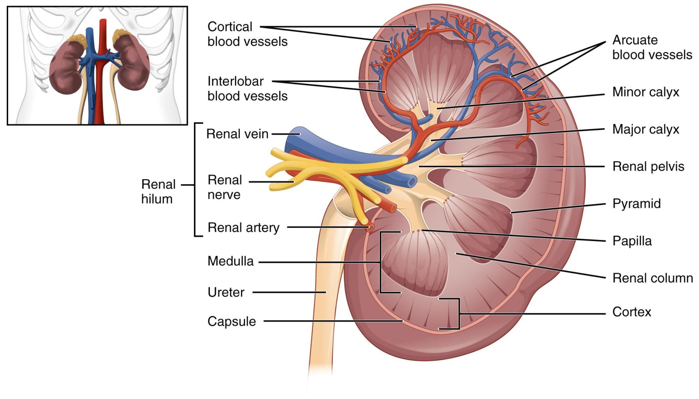
Cortical blood vessels: These are small arteries and veins that supply and drain blood from the renal cortex. They are crucial for delivering oxygen and nutrients to the cortical nephrons and for collecting filtered blood.
Interlobar blood vessels: These arteries and veins run between the renal pyramids in the renal columns. They branch off the renal artery and drain into the renal vein, providing blood supply to the renal lobes.
Renal vein: This large vein drains filtered, deoxygenated blood from the kidney back into the inferior vena cava. It carries blood that has had waste products removed.
Renal hilum: This is the concave indentation on the medial side of the kidney, serving as the entry and exit point for the renal artery, renal vein, ureter, and renal nerves. It is a critical anatomical landmark for kidney structure.
Renal nerve: These nerves, primarily sympathetic, innervate the kidney and play a role in regulating renal blood flow and renin secretion. They help control kidney function in response to systemic physiological demands.
Renal artery: This major artery branches directly from the aorta and supplies oxygenated blood to the kidney. It divides into smaller segmental arteries upon entering the hilum.
Medulla: This is the inner region of the kidney, characterized by the renal pyramids and columns. It contains structures crucial for concentrating urine, such as the loops of Henle and collecting ducts.
Ureter: This muscular tube emerges from the renal pelvis and carries urine from the kidney to the bladder. Peristaltic contractions ensure the unidirectional flow of urine.
Capsule: This tough, fibrous outer layer encases the kidney, providing protection against trauma and infection. It helps to maintain the kidney’s shape and integrity.
Arcuate blood vessels: These arteries and veins are found at the junction between the renal cortex and medulla, forming arches over the bases of the renal pyramids. They give rise to the cortical blood vessels.
Minor calyx: These cup-shaped structures collect urine from the renal papillae. Several minor calyces merge to form a major calyx.
Major calyx: These larger cup-shaped structures are formed by the convergence of several minor calyces. They further drain urine into the renal pelvis.
Renal pelvis: This funnel-shaped structure is located within the renal hilum, collecting urine from the major calyces. It acts as a reservoir before urine passes into the ureter.
Pyramid: Also known as renal pyramids, these cone-shaped tissues are located in the renal medulla. They contain segments of nephrons and collecting ducts, which are essential for urine formation and concentration.
Papilla: The apex of each renal pyramid, where urine drains into a minor calyx. It is perforated by collecting ducts that release urine.
Renal column: These extensions of cortical tissue project into the medulla, separating the renal pyramids. They contain blood vessels and provide structural support to the kidney.
Cortex: This is the outermost layer of the kidney, located just beneath the capsule. It contains the renal corpuscles and convoluted tubules of the nephrons, where the initial filtration of blood occurs.
The kidney is an extraordinarily efficient organ, responsible for a multitude of vital functions, including filtering blood, regulating blood pressure, maintaining electrolyte balance, and producing hormones. A detailed understanding of its internal architecture, as depicted in this sectional view, is fundamental to appreciating how these functions are accomplished. From the rich vascular supply entering through the renal hilum to the complex system of collecting ducts that funnel urine into the ureter, every part plays a specific, indispensable role. The image provides a clear roadmap of this intricate internal landscape, revealing the distinct regions and structures that collectively ensure the body’s internal environment remains stable.
The kidney’s functional unit, the nephron, spans both the cortex and medulla, illustrating the interdependent relationship between these regions. Blood enters the kidney via the renal artery, branching progressively into smaller arteries: segmental, interlobar, arcuate, and finally cortical arteries. These vessels deliver blood to the glomeruli within the renal corpuscles in the cortex, where the initial filtration of plasma occurs. The filtered fluid, now called filtrate, then passes through the renal tubules, which extend into the medulla within the renal pyramids. Here, essential substances are reabsorbed back into the blood, and additional waste products are secreted into the filtrate, precisely regulating the composition of urine.
As the filtrate travels through the various segments of the nephron and collecting ducts, its composition is continuously modified. The loops of Henle, located primarily in the medulla, are crucial for establishing the osmotic gradient necessary for concentrating urine. The collecting ducts, which receive filtrate from multiple nephrons, pass through the renal papillae and release the final urine into the minor calyces. These calyces then merge into major calyces, which in turn drain into the renal pelvis. From the renal pelvis, urine flows into the ureter and is transported to the bladder for storage, completing the journey. This highly organized structure ensures not only efficient waste removal but also the meticulous conservation of water and essential solutes, critical for maintaining overall physiological balance.
Disruptions to any part of this intricate system can lead to significant health problems. For instance, blockages in the renal artery can cause renal hypertension, while damage to the nephrons can lead to chronic kidney disease (CKD), affecting filtration and waste removal. Kidney stones, often forming in the calyces or renal pelvis, can obstruct urine flow, causing severe pain and potentially leading to kidney damage if not treated. Understanding the precise location and function of structures like the renal artery, renal vein, and the various calyces is therefore paramount for diagnosing and managing a wide array of renal pathologies, from infections to more complex systemic diseases impacting kidney health.

