Explore the fascinating microscopic world of a ray-finned fish vertebra, a testament to evolutionary design for aquatic locomotion. This article delves into the unique structure of these bony segments, revealing how each vertebra contributes to the fish’s flexibility and powerful swimming. Understand the biomechanical marvel that allows fish to navigate their watery environments with precision and speed.
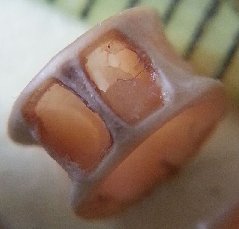
The vertebral column, the central axis of the skeletal system in vertebrates, exhibits remarkable diversity across different animal groups, reflecting adaptations to their specific environments and modes of life. In ray-finned fish, the most diverse group of vertebrates, the vertebrae are uniquely structured to facilitate efficient movement through water. Unlike the complex articulations seen in terrestrial vertebrates, fish vertebrae are often amphicoelous, meaning they are concave at both the anterior and posterior ends, forming hourglass-like shapes. This design, visible even in a small ray-finned fish vertebra, is crucial for both flexibility and the transfer of propulsive forces.
Each vertebra in a ray-finned fish is a compact, bony unit that contributes to the overall strength and elasticity of the spine. The concavities on either end of the vertebral body are typically filled with remnants of the notochord, a flexible rod that is a defining feature of chordates. This arrangement, along with the surrounding connective tissues, forms a continuous, flexible rod that allows the fish’s body to undulate in a wave-like motion, propelling it forward. The intricate internal structure of the vertebra provides strength while remaining lightweight, an essential balance for buoyancy and efficient swimming.
The vertebral column in fish serves multiple critical functions beyond mere structural support. It protects the delicate spinal cord, which runs through the neural canal of each vertebra, transmitting sensory and motor signals throughout the body. Furthermore, the numerous processes extending from each vertebra provide attachment sites for the powerful axial muscles, which are responsible for the undulatory swimming movements characteristic of most fish. The segmented nature of the spine, where each vertebra acts as a fulcrum for muscle action, allows for precise control over swimming direction and speed.
Key features of ray-finned fish vertebrae include:
- Amphicoelous centrum: Concave at both ends.
- Neural arch: Protects the spinal cord dorsally.
- Haemal arch: Protects blood vessels ventrally (in caudal vertebrae).
- Transverse processes: For muscle attachment.
- Notochord remnants: Provide flexibility between centra.
In conclusion, the vertebra of a small ray-finned fish, though seemingly simple, is a highly specialized anatomical structure perfectly engineered for an aquatic existence. Its amphicoelous design, in conjunction with the preserved notochordal material, allows for exceptional flexibility and efficient transmission of forces during undulatory swimming. This intricate skeletal component is fundamental to the fish’s ability to navigate its environment, escape predators, and hunt prey, showcasing the remarkable adaptations that have allowed ray-finned fish to dominate aquatic ecosystems across the globe.

