The female reproductive system is a marvel of biological design, centered around the interconnected functions of the ovaries, uterine tubes (fallopian tubes), and uterus. This detailed diagram provides a comprehensive anatomical overview, including microscopic insights into ovarian and uterine tissues. Understanding these vital organs and their intricate relationships is fundamental to comprehending female fertility, pregnancy, and overall reproductive health.
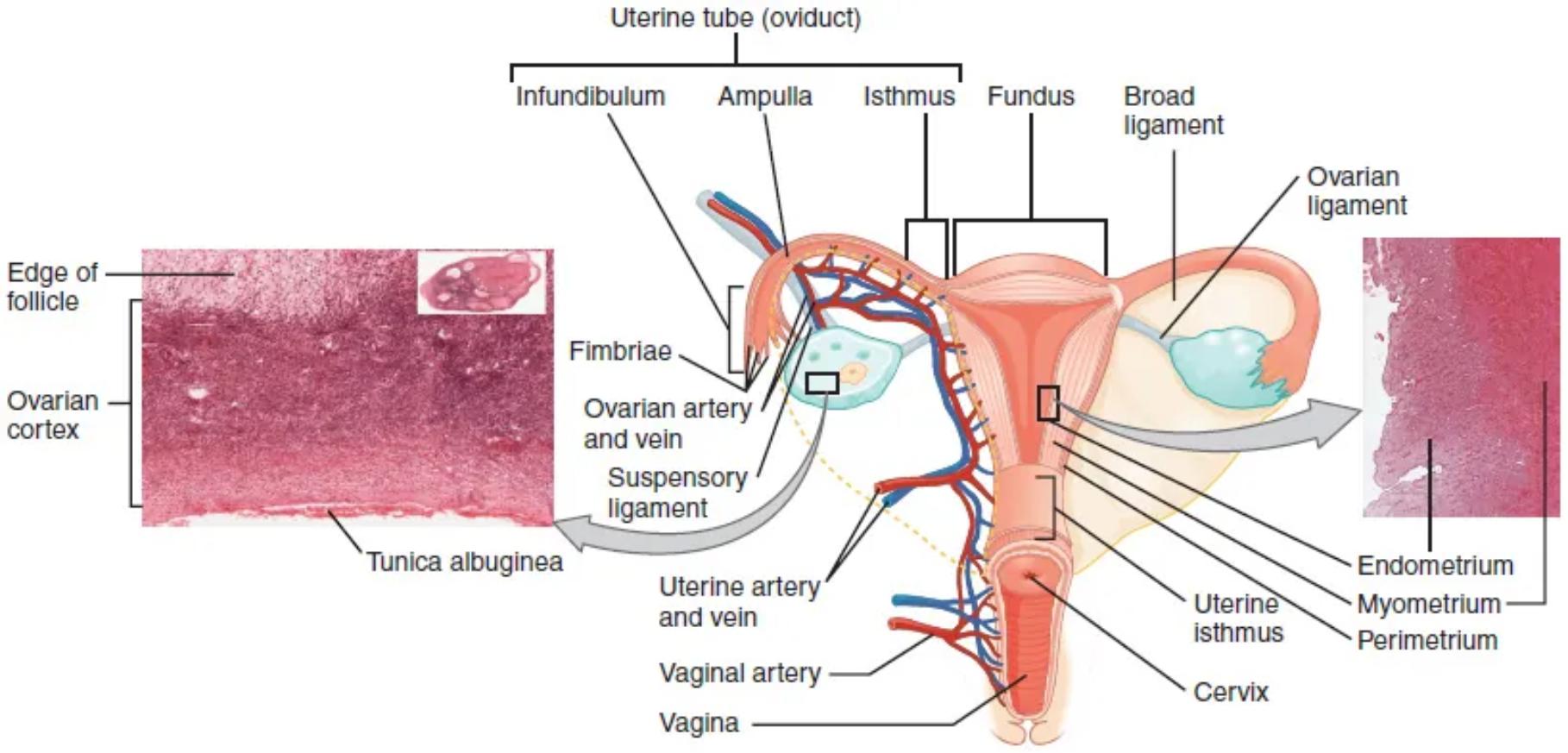
Uterine tube (oviduct): The uterine tube, also known as the fallopian tube or oviduct, is a muscular tube that extends from the uterus towards the ovary. It serves as the pathway for the ovum to travel from the ovary to the uterus and is the usual site of fertilization.
Infundibulum: The infundibulum is the funnel-shaped, most lateral part of the uterine tube, closest to the ovary. Its wide opening is fringed with fimbriae, which help to capture the ovum after ovulation.
Ampulla: The ampulla is the longest and widest part of the uterine tube, located medial to the infundibulum. This is typically the site where fertilization of the ovum by sperm occurs.
Isthmus: The isthmus is the narrow, constricted part of the uterine tube, connecting the ampulla to the uterus. Its muscular walls facilitate the movement of the fertilized egg towards the uterine cavity.
Fundus: The fundus is the dome-shaped, superior portion of the uterus, located above the entrance of the uterine tubes. It is the broadest part of the uterus and contributes significantly to uterine growth during pregnancy.
Broad ligament: The broad ligament is a wide, peritoneal fold that drapes over the uterus, uterine tubes, and ovaries, attaching them to the lateral walls of the pelvis. It provides support and contains the blood vessels, nerves, and lymphatic vessels supplying these organs.
Ovarian ligament: The ovarian ligament is a fibrous cord that connects the medial pole of the ovary to the lateral wall of the uterus, just below the uterine tube. It helps to anchor the ovary in its position within the pelvic cavity.
Fimbriae: Fimbriae are finger-like projections located at the free, distal end of the infundibulum of the uterine tube. Their sweeping movements create currents that help draw the released ovum into the uterine tube after ovulation.
Ovarian artery and vein: The ovarian artery and vein are the main blood vessels that supply and drain the ovaries, respectively. They travel within the suspensory ligament, providing essential nutrients and removing waste products from the ovarian tissues.
Suspensory ligament: The suspensory ligament is a fold of peritoneum that extends from the ovary to the lateral wall of the pelvis. It encases the ovarian artery, vein, and nerves, providing both support and a neurovascular pathway to the ovary.
Uterine artery and vein: The uterine artery and vein are the primary blood vessels supplying and draining the uterus, respectively. They are crucial for delivering oxygen and nutrients, especially to the metabolically active myometrium and endometrium.
Vaginal artery: The vaginal artery is a branch of the internal iliac artery that supplies blood to the vagina. It ensures adequate vascularization for the muscular wall and mucosal lining of the vagina.
Vagina: The vagina is a muscular, elastic tube extending from the cervix to the exterior of the body, forming the birth canal. It also serves as the receptacle for sperm during sexual intercourse and the pathway for menstrual flow.
Endometrium: The endometrium is the innermost lining of the uterus, a highly vascularized mucosal layer that undergoes cyclical changes in response to ovarian hormones. It is the site of embryo implantation and is shed during menstruation if pregnancy does not occur.
Myometrium: The myometrium is the thick, muscular middle layer of the uterine wall, composed primarily of smooth muscle fibers. Its contractions are responsible for expelling the fetus during childbirth and for menstrual cramps.
Perimetrium: The perimetrium is the outermost serous layer of the uterus, consisting of a thin layer of connective tissue covered by peritoneum. It forms part of the broad ligament and provides external protection to the uterus.
Uterine isthmus: The uterine isthmus is the narrowed, lower segment of the uterus, located between the body of the uterus (corpus) and the cervix. It is a transition zone that lengthens and thins during pregnancy.
Cervix: The cervix is the narrow, cylindrical lower portion of the uterus that projects into the vagina. It forms a canal that allows for the passage of sperm, menstrual blood, and a baby during childbirth, dilating significantly during labor.
Edge of follicle: This refers to the boundary of an ovarian follicle, which is the functional unit of the ovary containing an oocyte. Follicles develop in various stages within the ovarian cortex.
Ovarian cortex: The ovarian cortex is the outer, functional layer of the ovary where all ovarian follicles are located and develop. It is rich in connective tissue and houses millions of oocytes from birth.
Tunica albuginea: The tunica albuginea is a dense, white, fibrous connective tissue capsule that surrounds the entire ovary, lying beneath the outermost germinal epithelium. It provides structural integrity and protection to the ovarian tissue.
The Foundation of Female Reproduction
The female reproductive system is a remarkable and intricately designed biological network, with the ovaries, uterine tubes, and uterus forming its core functional unit. These organs work in concert to produce eggs, facilitate fertilization, and provide a nurturing environment for fetal development. The accompanying diagram offers a comprehensive anatomical depiction, detailing not only the macroscopic relationships but also microscopic insights into the tissues that comprise these vital structures. A thorough understanding of this anatomy is essential for grasping the complexities of human reproduction, gynecological health, and the mechanisms behind various reproductive conditions.
The reproductive process hinges on the precise coordination between these organs. The ovaries, as the primary female gonads, are responsible for gamete production and hormone synthesis. The uterine tubes act as conduits, capturing the ovum and serving as the typical site of fertilization. The uterus, a muscular marvel, is specialized for supporting and sustaining pregnancy. Together, these structures represent a finely tuned system, critical for the continuation of the human species.
Key components and their roles include:
- Ovaries: Egg production and hormone synthesis (estrogen, progesterone).
- Uterine tubes: Transport of ovum, site of fertilization.
- Uterus: Implantation of embryo, fetal development.
- Vagina: Birth canal, intercourse.
- Supporting ligaments and vascular supply.
Dysfunction in any part of this system can lead to significant reproductive health issues, underscoring the importance of its integrated functioning.
Anatomical Detail and Functional Interplay
The ovaries, typically almond-shaped organs, are strategically located in the pelvic cavity, anchored by the ovarian and suspensory ligaments. Their outer layer, the ovarian cortex, is where all the ovarian follicles reside and develop, from primordial stages to mature ovulatory follicles. The dense tunica albuginea provides a protective capsule. Adjacent to the ovaries are the uterine tubes, which are divided into distinct regions: the infundibulum with its fimbriae, responsible for capturing the ovum post-ovulation; the ampulla, where fertilization most commonly occurs; and the narrower isthmus, connecting to the uterus. The ciliated lining and muscular contractions of the uterine tubes facilitate the transport of the ovum or early embryo towards the uterus.
The uterus, a pear-shaped muscular organ, is centrally located in the pelvic cavity. Its most superior part is the fundus, while the main body is the corpus, which tapers down to the uterine isthmus and then the cervix. The uterine wall consists of three layers: the outer perimetrium, the thick muscular myometrium responsible for contractions during labor, and the inner endometrium. The endometrium is a highly dynamic layer that undergoes cyclical changes in response to ovarian hormones, thickening to prepare for embryo implantation and shedding during menstruation if pregnancy does not occur. A rich vascular network, including the ovarian and uterine arteries and veins, ensures that these organs receive adequate blood supply to support their high metabolic demands and endocrine functions. The vagina, a muscular and elastic tube, connects the uterus to the external environment, serving as the birth canal and for sexual intercourse. The intricate anatomical relationships and coordinated functions of these organs are paramount for female reproductive success.
In conclusion, the ovaries, uterine tubes, and uterus form the cornerstone of the female reproductive system, each playing distinct yet integrated roles. From the genesis of eggs and hormones in the ovaries to the intricate journey through the uterine tubes and the nurturing environment of the uterus, this system is a testament to biological complexity. A comprehensive understanding of their anatomy, vascular supply, and supporting structures is invaluable for medical professionals and for anyone seeking to deepen their knowledge of female health and the miraculous process of human reproduction.

