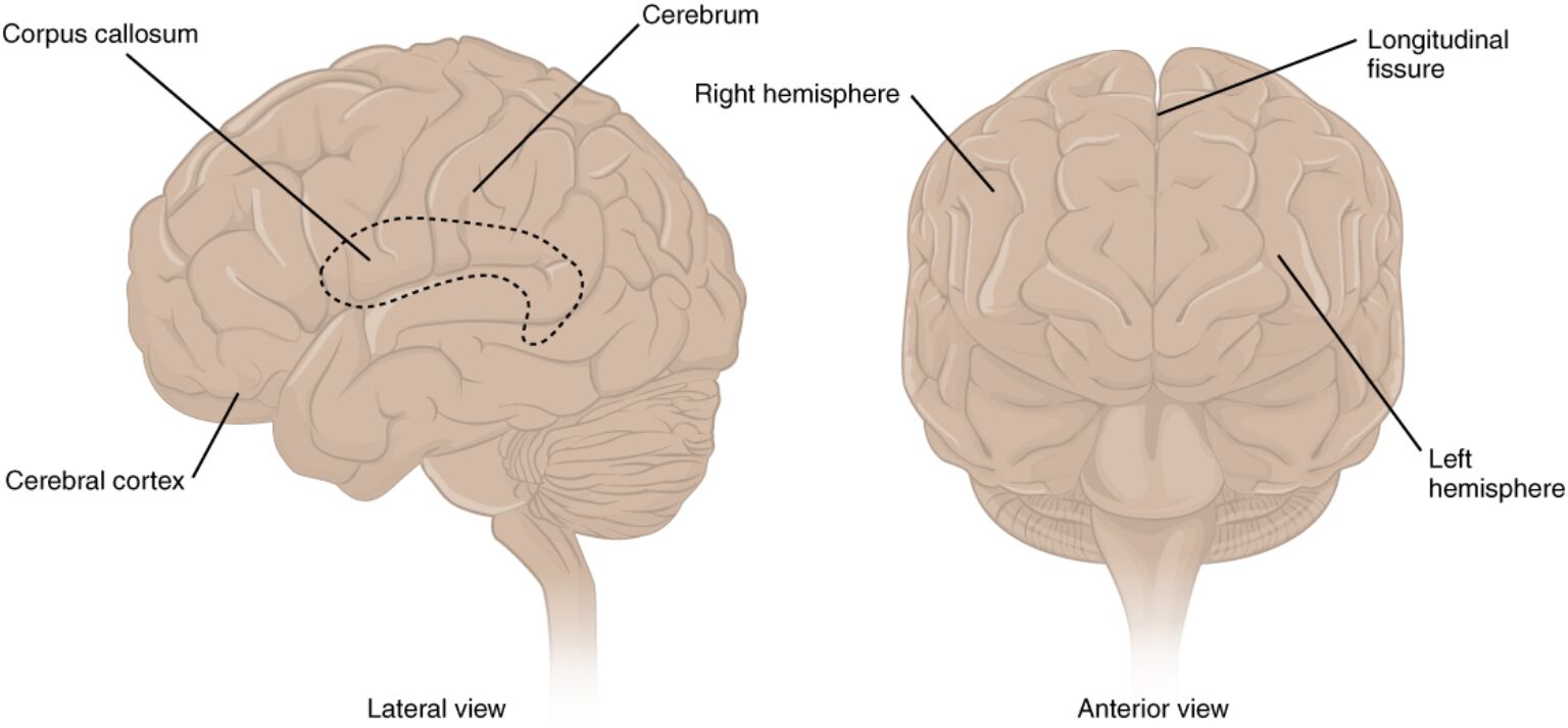
The cerebrum stands as the largest part of the human brain, dominating the central nervous system with its convoluted surface known as the cerebral cortex, which processes higher functions like thought, memory, and voluntary movement. This diagram presents lateral and anterior perspectives, labeling key features such as hemispheres, fissures, and connecting structures that facilitate interhemispheric communication and sensory-motor integration. By examining these views, one appreciates the cerebrum’s role in enabling complex behaviors unique to humans, from language to problem-solving.
Labeled Parts of the Cerebrum
Corpus callosum
The corpus callosum is a thick band of white matter fibers connecting the left and right hemispheres, visible in the lateral view as a curved structure beneath the cortex. It allows for the transfer of information between hemispheres, supporting coordinated functions like bilateral motor control and cognitive processing.
Cerebrum
The cerebrum encompasses the entire upper brain mass shown in both views, divided into lobes responsible for executive functions and perception. As the seat of consciousness, it integrates sensory inputs and generates outputs, with its size in humans reflecting advanced evolutionary development.
Right hemisphere
The right hemisphere appears on the left side of the anterior view, specializing in spatial awareness, creativity, and nonverbal communication. It processes holistic information, such as facial recognition, and collaborates with the left via the corpus callosum for unified brain activity.
Longitudinal fissure
The longitudinal fissure is the deep groove separating the two hemispheres, evident in the anterior view as a central divide. This fissure houses blood vessels and allows for independent yet interconnected hemispheric operations, crucial for lateralized functions like handedness.
Cerebral cortex
The cerebral cortex forms the outer folded layer of the cerebrum, labeled in the lateral view, consisting of gray matter rich in neuron cell bodies. Its gyri and sulci increase surface area for enhanced processing capacity, underpinning intelligence and adaptive behaviors.
Left hemisphere
The left hemisphere is shown on the right in the anterior view, typically dominant for language, logic, and analytical tasks in right-handed individuals. It handles sequential processing and communicates with the right hemisphere to integrate verbal and nonverbal cues.
Lateral view
The lateral view presents a side profile of the cerebrum, highlighting the corpus callosum and cerebral cortex’s contours. This perspective reveals the brain’s external features, aiding in understanding surface anatomy and lobe divisions.
Anterior view
The anterior view offers a front-facing look at the cerebrum, emphasizing the longitudinal fissure and hemispheric symmetry. It illustrates the brain’s bilateral organization, essential for studying asymmetries in function and pathology.
In-Depth Anatomy of the Cerebrum
The cerebrum’s structure is intricately designed for advanced neural processing, with its divided hemispheres and folded cortex maximizing computational power. Anatomical details reveal evolutionary optimizations for human cognition.
- The cerebral cortex, about 2-4 mm thick, contains six layers of neurons, with layer V pyramidal cells projecting to subcortical areas for motor output.
- Corpus callosum comprises over 200 million axons, divided into genu (frontal connections), body (parietal), and splenium (occipital), facilitating tasks like bimanual coordination.
- Hemispheres exhibit slight asymmetries, with the left often larger in language areas like Broca’s.
- Longitudinal fissure extends to the corpus callosum base, bordered by cingulate gyri involved in emotion.
- White matter beneath the cortex includes association fibers for intrahemispheric links and commissural for interhemispheric.
Physiological Functions of Cerebral Structures
Physiological roles center on integration and specialization, with the cortex processing vast inputs for output generation. Functions vary by region and hemisphere.
- The cerebral cortex handles sensory interpretation in parietal lobes and decision-making in frontal, with blood flow via middle cerebral artery supporting metabolism.
- Corpus callosum enables split-brain phenomena in sectioned patients, where hemispheres operate independently yet compensate.
- Right hemisphere excels in visuospatial tasks, processing global patterns via thalamic relays.
- Left hemisphere dominates analytical functions, with Wernicke’s area for comprehension linked to auditory cortex.
- Fissure separation allows lateralization, reducing redundancy while the callosum integrates for unified perception.
Hemispheric Specialization and Lateralization
Hemispheres exhibit functional asymmetries, shaped by genetics and experience. Lateralization enhances efficiency in processing.
- Language lateralizes to the left in 95% of right-handers, involving arcuate fasciculus connecting Broca’s and Wernicke’s areas.
- Right hemisphere manages prosody and emotional tone, with lesions causing aprosodia.
- Corpus callosum damage disrupts transfer, as in alexia without agraphia where right visual field info can’t reach left language centers.
- Developmental plasticity allows recovery from early lesions, with the opposite hemisphere assuming functions.
- Sex differences show females with thicker callosa, potentially aiding bilateral processing.
Developmental Biology of the Cerebrum
Cerebrum development begins in utero, with neural tube folding into prosencephalon. Processes involve proliferation and migration.
- Cortical neurogenesis peaks at weeks 12-16, with radial glia guiding neurons to layers.
- Callosum forms by week 20, axons crossing midline guided by slit-robo signaling.
- Hemispheric asymmetry emerges prenatally, influenced by genes like LRRTM1 for handedness.
- Thyroid hormones T3 and T4 regulate myelination of callosal fibers postnatally.
- Gyral formation by week 26 increases surface area, continuing into infancy.
Comparative and Evolutionary Perspectives
Comparing human cerebrum to other primates reveals expansions for cognition. Evolutionary changes emphasize adaptation.
- Human cortex surface area is 3 times larger than chimpanzees’, with more folds for neurons.
- Callosum relative size smaller in humans, suggesting greater hemispheric independence for specialization.
- Anterior view symmetry conserved across mammals, but human frontal enlargement supports planning.
- Fossil endocasts show gradual increase in hominid cortical complexity.
- Lateral view in apes shows less pronounced lobes, correlating with basic tool use vs. human innovation.
Research Techniques in Cerebral Anatomy
Methods visualize and probe cerebrum structures for research and diagnosis. Techniques advance understanding of function.
- fMRI maps activation in hemispheres during tasks, revealing lateralization.
- DTI traces callosal tracts, assessing integrity in multiple sclerosis.
- EEG records cortical rhythms, with alpha waves indicating relaxed states.
- Histology stains like Golgi reveal neuronal morphology in cortex layers.
- Optogenetics in models manipulates specific hemispheric circuits.
Clinical Significance and Pathologies
Cerebrum anomalies affect cognition and movement, with treatments targeting specific structures. Disorders often involve asymmetry or disconnection.
- Agenesis of corpus callosum leads to coordination issues, diagnosed via MRI and managed supportively.
- Stroke in left hemisphere causes aphasia, rehabilitated with speech therapy.
- Alzheimer’s cortical atrophy begins in temporal lobes, treated with cholinesterase inhibitors.
- Epilepsy from cortical malformations controlled by antiepileptics or surgery.
- Traumatic injuries to fissures cause hemorrhages, requiring neurosurgical intervention.
In summary, the diagram’s lateral and anterior views of the cerebrum illuminate its hemispheric division, cortical folds, and connecting corpus callosum, foundational to human neural architecture. This knowledge not only deepens appreciation of brain organization but also supports advancements in treating cerebral disorders, enhancing neurological care.

