Explore the fundamental processes of systole and diastole, the two critical phases that define the heart’s rhythmic action. This article delves into how the heart efficiently pumps blood to the body during systole and replenishes its chambers during diastole, highlighting the importance of each phase for cardiovascular health. Understand the coordinated muscle contractions and relaxations that ensure continuous blood circulation, a vital function for sustaining life.
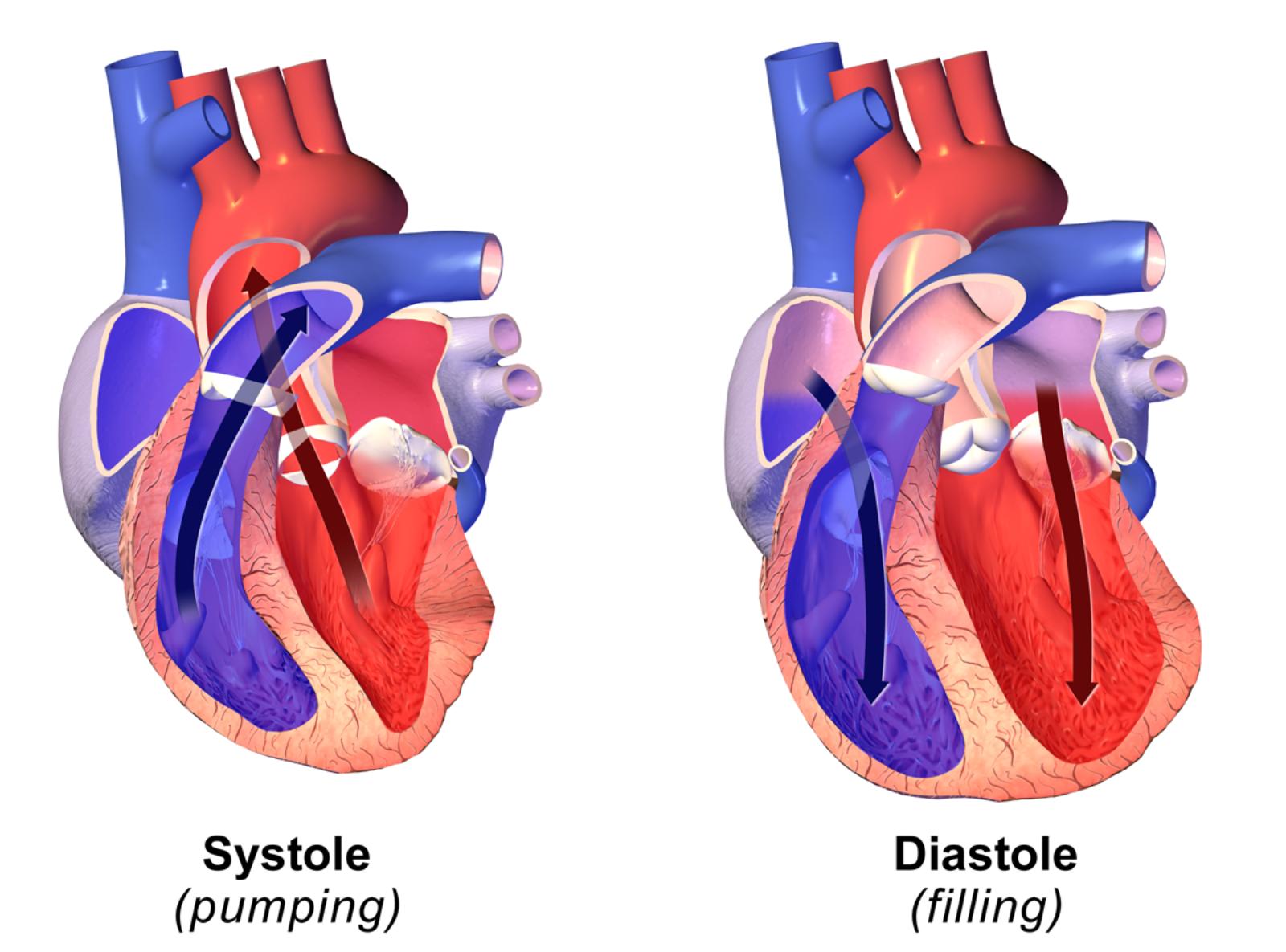
Systole (pumping): This label refers to the contraction phase of the cardiac cycle, specifically ventricular systole. During this period, the heart’s ventricles contract forcefully to eject blood into the major arteries. This action is crucial for distributing oxygenated blood to the body and deoxygenated blood to the lungs.
Diastole (filling): This label represents the relaxation phase of the cardiac cycle, known as ventricular diastole. In this phase, the heart’s ventricles relax and expand, allowing them to fill with blood from the atria. Proper filling during diastole is essential for ensuring that enough blood is available for the next systolic contraction.
The human heart is a remarkable organ, continuously working to circulate blood throughout the body. This vital process is orchestrated by a precise, rhythmic cycle of contraction and relaxation, known as the cardiac cycle. The image visually distinguishes between the two primary phases of this cycle: systole, the pumping phase, and diastole, the filling phase. Together, these two actions ensure that every cell in the body receives the oxygen and nutrients it needs to function, while waste products are efficiently removed.
Understanding systole and diastole is fundamental to comprehending cardiovascular physiology and many heart conditions. During systole, the muscular walls of the heart, particularly the ventricles, contract to generate the pressure required to propel blood into the arteries. Conversely, diastole is a period of relaxation and refilling, where the heart chambers expand to draw blood in. The seamless transition between these two phases is vital for maintaining blood pressure and cardiac output.
The visual representation clearly illustrates the direction of blood flow during each phase. In systole, arrows indicate blood being forcefully ejected from the ventric into the great arteries. In diastole, arrows show blood flowing from the atria into the relaxed ventricles, preparing for the subsequent contraction. This cyclical pumping action is not only continuous but also highly adaptable, adjusting to the body’s varying demands for blood supply.
- Systole is the heart’s pumping phase.
- Diastole is the heart’s filling phase.
- These phases regulate blood pressure.
- Proper function is crucial for cardiac output.
The Dynamics of Systole: Pumping Life Through the Body
Systole, derived from the Greek word meaning “contraction,” represents the active pumping phase of the cardiac cycle. During this period, the ventricles of the heart contract vigorously, increasing pressure within their chambers. This pressure rise forces open the semilunar valves (aortic and pulmonic valves) and propels blood into the aorta and pulmonary artery, respectively. The left ventricle, being the stronger of the two, generates significantly higher pressure to pump oxygenated blood throughout the entire systemic circulation, reaching every tissue and organ. Simultaneously, the right ventricle pumps deoxygenated blood to the lungs via the pulmonary artery for oxygenation. The duration of systole is typically shorter than diastole, yet it is the primary force responsible for maintaining blood pressure and tissue perfusion. Any abnormality in systolic function, such as in heart failure with reduced ejection fraction, can significantly impair the body’s ability to receive adequate blood supply, leading to symptoms like fatigue and shortness of breath.
The Importance of Diastole: Relaxing and Refilling for the Next Beat
Diastole, from the Greek word for “dilation,” is the essential relaxation and filling phase of the heart. During this period, the ventricular muscles relax, causing the pressure within the chambers to drop. This pressure decrease allows the atrioventricular valves (mitral and tricuspid valves) to open, facilitating the flow of blood from the atria into the ventricles. Approximately 70-80% of ventricular filling occurs passively during early diastole, with the remaining 20-30% contributed by atrial contraction (the “atrial kick”) at the very end of diastole. Adequate diastolic filling is critical because it determines the amount of blood available for the next systolic ejection—a concept known as preload. Conditions like diastolic dysfunction, often seen in hypertension or hypertrophic cardiomyopathy, impair the heart’s ability to relax and fill properly, leading to reduced cardiac output despite normal systolic function.
The synchronized interplay between systole and diastole is a testament to the intricate engineering of the human heart. Each phase is interdependent, with the efficiency of one directly influencing the other. Maintaining a healthy balance and proper function of both pumping and filling mechanisms is paramount for overall cardiovascular health. Regular monitoring of blood pressure, which reflects both systolic and diastolic pressures, provides crucial insights into the heart’s performance and can indicate underlying issues that require medical attention. This fundamental understanding of systole and diastole is a cornerstone in preventing and managing heart disease.

