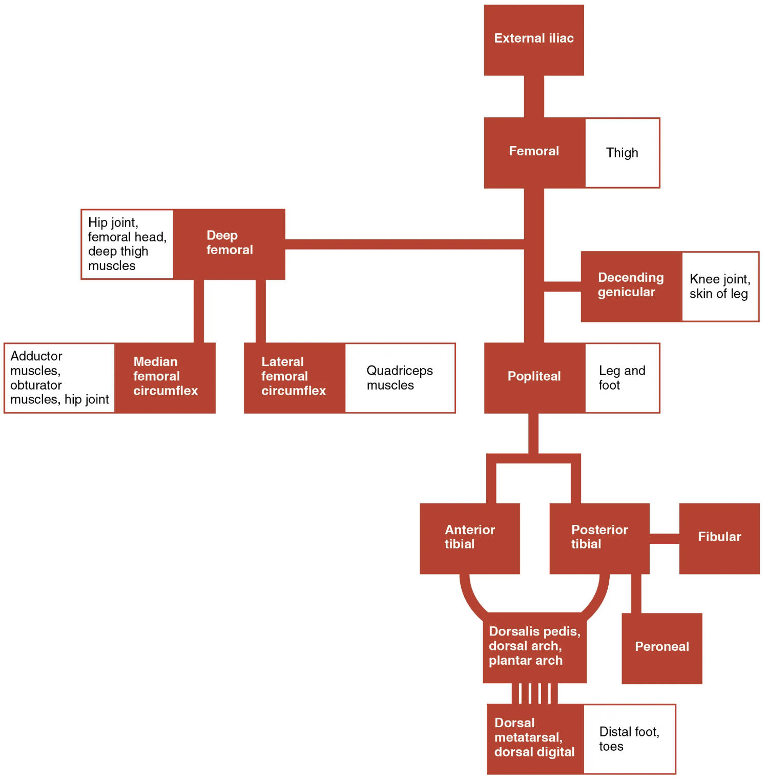
The lower limb’s systemic arteries play a crucial role in delivering oxygenated blood from the heart to support movement, muscle function, and tissue health. This flowchart illustrates the distribution of arteries starting from the external iliac artery, branching extensively to nourish the thigh, leg, and foot, providing an essential resource for understanding circulatory anatomy.
Exploring the Key Arteries in the Flowchart
The labeled arteries in this image outline the systemic circulation to the lower limb, originating from the external iliac artery. Each vessel contributes uniquely to the vascular network, ensuring comprehensive blood supply.
External iliac The external iliac artery branches from the common iliac, transitioning into the femoral artery below the inguinal ligament. It serves as the primary conduit for blood flow to the lower limb, supporting pelvic and thigh circulation.
Femoral The femoral artery, a continuation of the external iliac, runs through the thigh, supplying the quadriceps and other muscles. It acts as a major artery for the upper leg and is a key site for pulse assessment.
Thigh The thigh region receives blood from the femoral artery, nourishing the surrounding muscles and tissues. This area includes branches that support the thigh’s extensive muscular structure.
Deep femoral The deep femoral artery branches from the femoral, supplying the deep thigh muscles and hip joint. It provides a robust collateral network, enhancing blood flow to the posterior thigh.
Hip joint, femoral head, deep thigh muscles The hip joint, femoral head, deep thigh muscles are supplied by branches of the deep femoral, ensuring joint stability and muscle function. These vessels support weight-bearing and hip movement.
Adductor muscles, muscles, hip joint The adductor muscles, muscles, hip joint receive blood from the deep femoral’s median circumflex branch, nourishing the inner thigh and hip. This supply is vital for adduction and hip stability.
Median femoral circumflex The median femoral circumflex artery, a branch of the deep femoral, supplies the medial thigh muscles and hip joint. It contributes to the anastomotic network around the hip, enhancing circulation.
Lateral femoral circumflex The lateral femoral circumflex artery branches from the deep femoral, supplying the lateral thigh muscles. It supports hip abduction and contributes to thigh stability.
Quadriceps muscles The quadriceps muscles receive blood from the femoral artery, ensuring strength for knee extension. This supply is critical for activities like walking and running.
Descending genicular The descending genicular artery branches from the femoral, supplying the knee joint and skin of the leg. It forms part of the genicular anastomosis, supporting knee function.
Knee joint, skin of leg The knee joint, skin of leg are nourished by the descending genicular artery, ensuring joint mobility and skin integrity. This supply aids in healing and joint stability.
Popliteal The popliteal artery continues from the femoral behind the knee, supplying the leg and foot. It bifurcates into the anterior and posterior tibial arteries, critical for lower leg circulation.
Leg and foot The leg and foot receive blood from the popliteal artery, supporting the entire lower limb distal to the knee. This extensive supply ensures mobility and tissue health.
Anterior tibial The anterior tibial artery branches from the popliteal, running along the shin to supply the anterior leg muscles. It continues as the dorsalis pedis, feeding the foot’s dorsal surface.
Posterior tibial The posterior tibial artery, a popliteal branch, runs along the calf, supplying the posterior muscles and foot sole. It contributes to the plantar arch, ensuring foot perfusion.
Fibular The fibular artery branches from the posterior tibial, supplying the lateral leg. It provides collateral circulation, supporting the fibular muscles and ankle stability.
Dorsalis pedis, dorsal arch, plantar arch The dorsalis pedis, dorsal arch, plantar arch system, originating from the anterior tibial, supplies the foot’s dorsal and plantar surfaces. This network ensures comprehensive foot circulation.
Peroneal The peroneal artery, another posterior tibial branch, supplies the lateral leg and ankle. It enhances blood flow to the peroneal region, supporting lateral stability.
Dorsal metatarsal, dorsal digital The dorsal metatarsal, dorsal digital arteries branch from the dorsalis pedis, supplying the metatarsals and toes. They ensure blood flow to the foot’s dorsal digits and bones.
Distal foot, toes The distal foot, toes receive blood from the dorsal and plantar arches, supporting the toes and distal foot. This supply is vital for sensation and movement.
The Pathway of Systemic Arteries
Blood flow to the lower limb begins with the external iliac artery, following a structured route through the thigh and leg. This pathway adapts to the limb’s anatomical and functional needs.
- The external iliac artery transitions into the femoral artery, initiating thigh circulation.
- The deep femoral artery branches to supply the hip and thigh muscles.
- The popliteal artery behind the knee splits into anterior and posterior tibial arteries.
- Distal arteries like the dorsalis pedis and plantar arch ensure foot and toe perfusion.
Clinical Relevance of Lower Limb Arteries
Understanding these arteries aids in diagnosing vascular conditions. Their positions guide clinical assessments and interventions.
- The femoral artery is a primary site for arterial access and pulse monitoring.
- The popliteal artery is assessed for aneurysms due to its posterior location.
- Genicular arteries support knee surgeries with collateral flow.
- Doppler ultrasound evaluates flow in the tibial and peroneal arteries.
Physiological Roles in Lower Limb Function
These arteries adapt to physical demands, ensuring oxygen delivery. Their responsiveness enhances mobility and endurance.
- The femoral and deep femoral arteries dilate during exercise, boosting thigh performance.
- The dorsal and plantar arches regulate blood flow for standing and walking.
- Hormonal influences, like adrenaline, modulate vascular tone in the lower limb.
- Anastomoses around the knee protect against ischemic events.
In conclusion, the systemic arteries of the lower limb form a resilient network supporting movement and stability. Mastering this anatomy enhances clinical practice and deepens appreciation for circulatory efficiency.

