The systemic arteries form a critical network that distributes oxygenated blood from the heart to every part of the body, sustaining life and function. This diagram highlights the major arteries, showcasing their pathways and roles in delivering nutrients and oxygen to tissues while removing waste products.
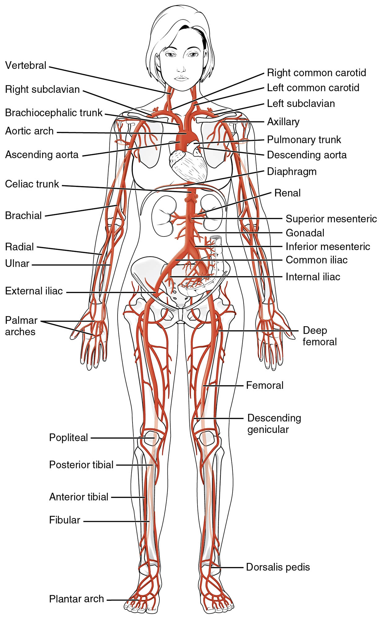
Aorta The aorta is the largest artery, originating from the left ventricle to carry oxygenated blood. It branches into smaller arteries to supply the entire body with vital nutrients.
Coronary arteries These arteries supply oxygenated blood to the heart muscle itself. They ensure the myocardium receives adequate oxygen for continuous pumping action.
Brachiocephalic artery This artery arises from the aorta and splits to supply the right arm and head. It delivers blood to the right subclavian and right common carotid arteries.
Left common carotid artery Originating from the aortic arch, it provides blood to the left side of the head and neck. It ensures oxygen delivery to the brain and facial structures.
Left subclavian artery This artery branches from the aortic arch to supply the left arm. It supports the upper limb with oxygenated blood for movement and function.
Celiac trunk Emerging from the abdominal aorta, it supplies blood to the stomach, liver, and spleen. It plays a key role in digesting nutrients and detoxifying blood.
Superior mesenteric artery This artery arises from the abdominal aorta to feed the small intestine and part of the large intestine. It ensures nutrient absorption and gastrointestinal health.
Renal arteries These arteries branch from the abdominal aorta to supply the kidneys. They deliver blood for filtration and regulation of blood pressure and volume.
Inferior mesenteric artery Originating from the abdominal aorta, it supplies the lower large intestine. It supports digestion and waste elimination in the distal colon.
Common iliac arteries These arteries split from the abdominal aorta to supply the pelvis and lower limbs. They ensure blood flow to the legs and reproductive organs.
Femoral arteries Continuing from the iliac arteries, they provide blood to the thighs and legs. They support muscle function and lower limb circulation.
Anatomy of the Systemic Arteries
The aorta serves as the starting point of the systemic arterial system, distributing blood efficiently. Its structure and branching pattern are designed for high-pressure flow.
- The ascending aorta rises from the heart, giving rise to the coronary arteries.
- The aortic arch curves, producing the brachiocephalic, left common carotid, and left subclavian arteries.
- The descending thoracic aorta continues to supply the chest and upper abdomen.
- The abdominal aorta further branches to reach visceral and lower body organs.
- Elastic fibers in its walls help maintain steady blood flow during systole and diastole.
Key Branches and Their Functions
The coronary arteries and other major branches play specific roles in oxygen delivery. Each artery targets critical regions to support bodily functions.
- Coronary arteries nourish the heart, preventing ischemia during activity.
- The brachiocephalic artery ensures blood reaches the brain and right arm.
- The left common carotid and subclavian arteries support left-side circulation.
- The celiac trunk feeds digestive organs, aiding metabolism and detoxification.
- These branches adapt to varying demands, such as during exercise.
Abdominal and Lower Body Arteries
The renal arteries and lower body arteries like the femoral arteries are essential for visceral and limb perfusion. Their roles extend beyond simple delivery.
- Renal arteries supply the kidneys, enabling filtration and hormone production.
- The superior and inferior mesenteric arteries nourish the intestines.
- Common iliac arteries branch to support pelvic and leg circulation.
- Femoral arteries ensure robust blood flow to active leg muscles.
- These arteries adjust flow based on metabolic needs and posture.
Physiological Importance of Systemic Circulation
The systemic arteries maintain blood pressure and nutrient distribution throughout the body. Their function is critical for overall homeostasis.
- The aorta’s elasticity smooths out pressure waves from the heart.
- Arterial branching reduces resistance, optimizing tissue perfusion.
- Oxygenated blood supports cellular respiration and energy production.
- Waste products like carbon dioxide are carried away for excretion.
- This system’s integrity is vital for preventing hypoxia or tissue damage.
Clinical Relevance of Arterial Anatomy
Knowledge of systemic artery anatomy aids in diagnosing and treating vascular conditions. The layout guides medical interventions effectively.
- Blockages in coronary arteries can lead to myocardial infarction.
- Aneurysms in the aorta require surgical monitoring or repair.
- Renal arteries issues may cause hypertension or kidney dysfunction.
- Femoral artery disease affects lower limb mobility and healing.
- Imaging techniques like angiography map these arteries for treatment planning.
The systemic arteries, led by the aorta, form a remarkable network that delivers oxygenated blood to sustain every tissue. Their intricate branching and adaptive responses ensure efficient circulation, providing a foundation for understanding cardiovascular health and addressing related challenges.

