The posterior aspect of the lower leg contains a vital group of muscles that underpin key movements and stability of the foot and ankle. This article examines the superficial muscles of the right lower leg, depicted in a posterior view, offering a detailed exploration of their anatomical structure and functional roles. These muscles, primarily located in the posterior compartment, are responsible for plantar flexion, with some contributing to inversion, eversion, and rotation of the foot, enhancing overall lower limb mobility. Through the labeled diagram, readers can develop a thorough understanding of these muscles’ significance in leg function and clinical applications.
Introduction to the Superficial Muscles of the Lower Leg
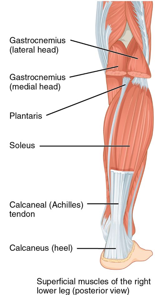
The superficial muscles of the right lower leg form the outer layer of the posterior compartment. Their posterior view highlights their critical role in foot and leg movement. This section details the labeled structures that define their anatomy and purpose.
- Gastrocnemius (lateral head): Positioned on the outer calf, this muscle plantar flexes the foot and flexes the knee. It provides significant power during walking or jumping.
- Gastrocnemius (medial head): Located on the inner calf, it plantar flexes the foot and flexes the knee. It works with the lateral head to strengthen the calf.
- Plantaris: A small muscle near the gastrocnemius, it weakly plantar flexes the foot. It assists in knee flexion and supports the Achilles tendon.
- Soleus: Found deep to the gastrocnemius, this muscle plantar flexes the foot. It is essential for maintaining posture and standing stability.
- Calcaneal (Achilles) tendon: Connects the calf muscles to the heel, it transmits force for plantar flexion. It is crucial for walking, running, and jumping.
- Calcaneus (heel): The heel bone serves as an insertion point for the Achilles tendon. It anchors the posterior muscles for effective movement.
The superficial muscles of the right lower leg‘s posterior placement ensures robust support. Their labeled view provides a clear understanding of their structural and functional contributions.
Functional Roles of the Superficial Muscles
The superficial muscles of the right lower leg are key to powerful foot movements. Their actions in the posterior compartment support stability and propulsion. This section outlines their specific functional roles.
- The gastrocnemius (lateral head) and gastrocnemius (medial head) plantar flex the foot. They also flex the knee, aiding in the push-off phase of gait.
- The plantaris weakly plantar flexes the foot, providing minor support. It assists in knee flexion, contributing to overall leg flexibility.
- The soleus plantar flexes the foot, maintaining posture during standing. It works continuously to support body weight over time.
- The calcaneal tendon transmits force from the calf muscles to the heel. This action powers plantar flexion during dynamic movements.
- The calcaneus serves as a stable base for muscle insertion. Its role enhances the efficiency of posterior muscle actions.
The superficial muscles of the right lower leg‘s coordinated efforts optimize foot function. Their posterior focus ensures effective movement and stability.
Clinical Significance and Practical Applications
The superficial muscles of the right lower leg are often assessed in clinical evaluations of leg and foot health. Their condition directly impacts mobility and daily activities. This section explores their clinical relevance.
- Strain in the gastrocnemius can lead to calf tightness or Achilles tendonitis. Stretching and strengthening exercises help restore flexibility and strength.
- Weakness in the soleus may cause difficulty with prolonged standing. Targeted training improves endurance and posture support.
- Injury to the calcaneal tendon can result in a ruptured Achilles, affecting plantar flexion. Surgical or conservative treatment may be required for recovery.
- Overuse of the plantaris may contribute to posterior leg pain. Rest and conditioning alleviate discomfort and prevent further issues.
- Understanding their anatomy aids in diagnosing conditions like calf muscle strain. This knowledge guides effective treatment and prevention strategies.
This insight is valuable for professionals addressing leg concerns. The superficial muscles of the right lower leg‘s roles highlight the need for precise therapeutic interventions.
Conclusion
The superficial muscles of the right lower leg, as illustrated in the posterior view, showcase the intricate design supporting foot and leg mobility. This article has explored their anatomical structure, diverse functional roles, and clinical significance, providing a thorough understanding of their importance. From the gastrocnemius powering plantar flexion to the soleus maintaining posture, each muscle contributes uniquely to lower limb stability and movement. Continued study of these muscles will enhance therapeutic approaches and deepen appreciation for the complex mechanics of the lower leg.

