The lower leg’s functionality hinges on a well-coordinated set of muscles that enable essential movements for daily activities. This article explores the superficial muscles of the right lower leg, presented in an anterior view, to provide a detailed examination of their anatomical structure and roles. These muscles, primarily located in the anterior compartment, are responsible for dorsiflexion, while lateral muscles assist in eversion and rotation of the foot, contributing to overall leg stability and mobility. Through the labeled diagram, readers can gain a comprehensive understanding of these muscles’ importance in foot and leg function.
Introduction to the Superficial Muscles of the Lower Leg
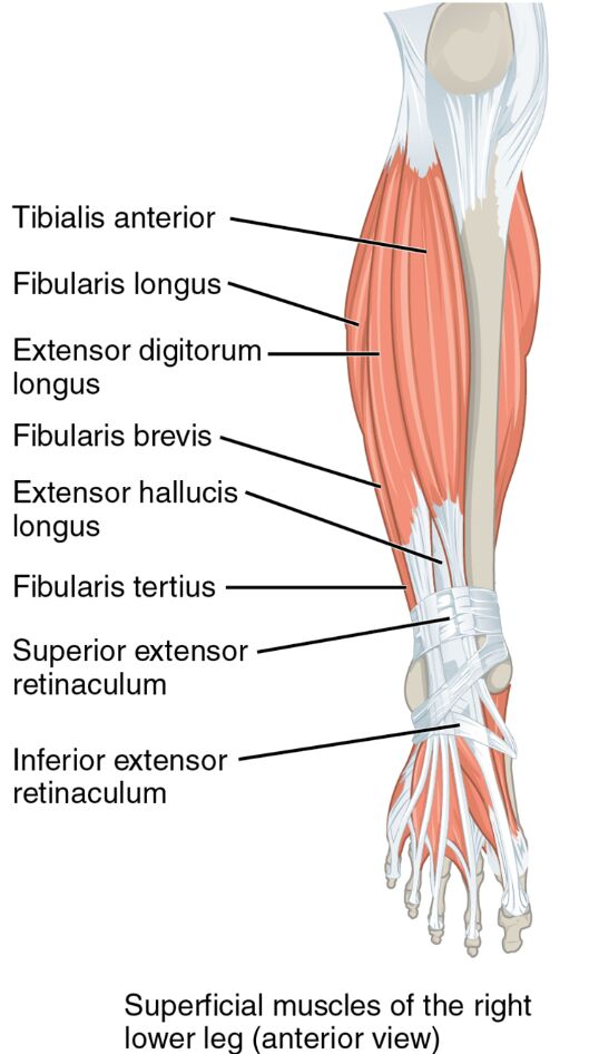
The superficial muscles of the right lower leg form the outer layer of the anterior compartment. Their visible arrangement supports critical leg movements from the front. This section details the labeled structures that define their anatomy and function.
- Tibialis anterior: Positioned on the front of the shin, this muscle dorsiflexes and inverts the foot. It lifts the foot to prevent tripping during walking.
- Fibularis longus: Located on the lateral side, this muscle everts and plantar flexes the foot. It enhances stability on uneven terrain.
- Extensor digitorum longus: Found along the anterior lateral shin, it extends the toes and dorsiflexes the foot. It aids in toe lifting during gait.
- Fibularis brevis: Positioned laterally below the fibularis longus, it everts the foot and assists in plantar flexion. It supports lateral ankle stability.
- Extensor hallucis longus: Located deeper in the anterior compartment, it extends the big toe and dorsiflexes the foot. It is essential for precise toe movements.
- Fibularis tertius: Found on the anterior lateral side, it dorsiflexes and everts the foot. It assists in foot elevation during motion.
- Superior extensor retinaculum: A band on the anterior ankle, it holds extensor tendons in place. It ensures smooth tendon movement during dorsiflexion.
- Inferior extensor retinaculum: Another anterior band, it stabilizes extensor tendons. It prevents tendon displacement during foot motion.
The superficial muscles of the right lower leg‘s anterior placement optimizes leg function. Their labeled view offers a clear insight into their structural and operational roles.
Functional Roles of the Superficial Muscles
The superficial muscles of the right lower leg are vital for specific movements of the foot and ankle. Their actions support both stability and mobility in the lower limb. This section outlines their functional contributions.
- The tibialis anterior dorsiflexes the foot, lifting it during the swing phase of walking. It also inverts the foot, aiding in maintaining arch support.
- The fibularis longus and fibularis brevis evert the foot, providing lateral stability. They assist in plantar flexion, enhancing push-off strength.
- The extensor digitorum longus extends the toes, facilitating toe lifting. It also dorsiflexes the foot, supporting smooth gait transitions.
- The extensor hallucis longus extends the big toe, improving toe-off precision. It contributes to dorsiflexion, aiding in foot clearance.
- The fibularis tertius dorsiflexes and everts the foot, enhancing foot elevation. It supports lateral movement and balance on uneven surfaces.
- The superior extensor retinaculum and inferior extensor retinaculum stabilize the extensor tendons. They ensure efficient force transmission during movement.
The superficial muscles of the right lower leg‘s coordinated actions enhance leg performance. Their specific roles ensure effective foot and ankle motion.
Clinical Significance and Practical Applications
The superficial muscles of the right lower leg are often evaluated in clinical assessments of leg and foot function. Their health directly influences mobility and daily activities. This section explores their clinical importance.
- Weakness in the tibialis anterior can lead to foot drop, causing tripping risks. Targeted exercises strengthen this muscle to improve dorsiflexion.
- Strain in the fibularis longus may cause ankle instability or eversion injuries. Rehabilitation focuses on restoring lateral support and flexibility.
- Overuse of the extensor digitorum longus can result in tendonitis, affecting toe extension. Rest and stretching alleviate pain and prevent further damage.
- Injury to the extensor hallucis longus may impair big toe movement. Physical therapy helps regain precision and dorsiflexion strength.
- Understanding their anatomy aids in diagnosing conditions like anterior compartment syndrome. This knowledge guides effective treatment and preventive measures.
This insight is valuable for professionals addressing leg issues. The superficial muscles of the right lower leg‘s roles underscore the need for precise therapeutic interventions.
Conclusion
The superficial muscles of the right lower leg, as depicted in the anterior view, highlight the intricate design supporting leg and foot mobility. This article has explored their anatomical structure, diverse functional roles, and clinical significance, providing a thorough understanding of their importance. From the tibialis anterior enabling dorsiflexion to the fibularis longus enhancing eversion, each muscle contributes uniquely to lower limb stability and movement. Continued study of these muscles will enhance therapeutic strategies and deepen appreciation for the complex mechanics of the lower leg.

