The lower leg’s superficial muscles are integral to the mobility and stability of the foot and ankle, forming a dynamic network essential for daily activities. This article investigates the superficial muscles of the right lower leg, presented in both anterior and posterior views, to offer a detailed exploration of their anatomical structure and functional significance. These muscles, primarily responsible for dorsiflexion in the anterior compartment and plantar flexion in the posterior compartment, also contribute to inversion, eversion, and rotation of the foot through lateral and medial actions. By examining the labeled diagrams, readers can gain a comprehensive understanding of these muscles’ roles in leg function and their relevance in clinical practice.
Introduction to the Superficial Muscles of the Lower Leg
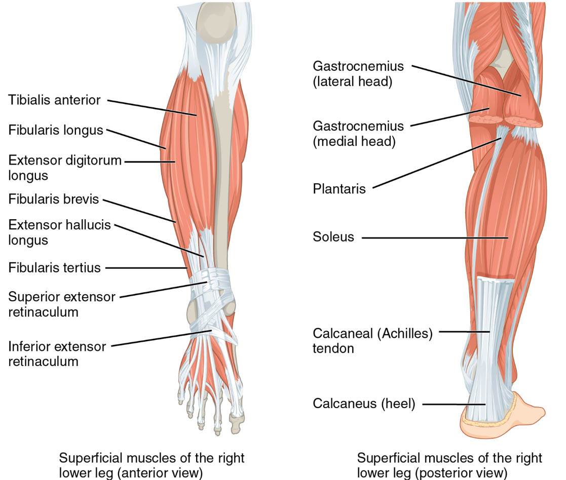
The superficial muscles of the right lower leg provide the outer muscular layer across both anterior and posterior views. Their dual perspectives highlight their diverse roles in leg movement. This section details the labeled structures that define their anatomy and function.
- Tibialis anterior: Positioned on the anterior shin, this muscle dorsiflexes and inverts the foot. It lifts the foot to prevent tripping during walking.
- Fibularis longus: Located on the lateral side in the anterior view, it everts and plantar flexes the foot. It enhances stability on uneven terrain.
- Extensor digitorum longus: Found along the anterior lateral shin, it extends the toes and dorsiflexes the foot. It aids in toe lifting during gait.
- Fibularis brevis: Positioned laterally in the anterior view, it everts the foot and assists in plantar flexion. It supports lateral ankle stability.
- Extensor hallucis longus: Located deeper in the anterior compartment, it extends the big toe and dorsiflexes the foot. It is essential for precise toe movements.
- Fibularis tertius: Found on the anterior lateral side, it dorsiflexes and everts the foot. It assists in foot elevation during motion.
- Superior extensor retinaculum: A band on the anterior ankle, it holds extensor tendons in place. It ensures smooth tendon movement during dorsiflexion.
- Inferior extensor retinaculum: Another anterior band, it stabilizes extensor tendons. It prevents tendon displacement during foot motion.
- Gastrocnemius (lateral head): Positioned on the outer calf in the posterior view, it plantar flexes the foot and flexes the knee. It provides significant power during walking.
- Gastrocnemius (medial head): Located on the inner calf in the posterior view, it plantar flexes the foot and flexes the knee. It works with the lateral head to strengthen the calf.
- Plantaris: A small muscle near the gastrocnemius in the posterior view, it weakly plantar flexes the foot. It assists in knee flexion and supports the Achilles tendon.
- Soleus: Found deep to the gastrocnemius in the posterior view, it plantar flexes the foot. It is vital for maintaining posture and standing stability.
- Calcaneal (Achilles) tendon: Connects the calf muscles to the heel in the posterior view, it transmits force for plantar flexion. It is crucial for walking, running, and jumping.
- Calcaneus (heel): The heel bone in the posterior view serves as an insertion point for the Achilles tendon. It anchors the posterior muscles for effective movement.
The superficial muscles of the right lower leg‘s dual views reveal their comprehensive support. Their labeled anatomy provides a foundation for understanding leg dynamics.
Functional Roles of the Superficial Muscles
The superficial muscles of the right lower leg are essential for a range of foot and ankle movements. Their actions across anterior and posterior compartments enhance mobility and stability. This section outlines their specific functional contributions.
- The tibialis anterior and extensor hallucis longus dorsiflex the foot in the anterior view. They lift the foot, preventing tripping and aiding in smooth gait.
- The fibularis longus and fibularis brevis evert the foot in the anterior view. They provide lateral stability, supporting balance on uneven surfaces.
- The extensor digitorum longus extends the toes and dorsiflexes the foot. This action facilitates toe lifting and foot clearance during walking.
- The gastrocnemius (lateral head) and gastrocnemius (medial head) plantar flex the foot in the posterior view. They also flex the knee, powering the push-off phase.
- The soleus plantar flexes the foot in the posterior view, maintaining posture. It supports prolonged standing by stabilizing the leg.
- The calcaneal tendon transmits force for plantar flexion in the posterior view. It ensures efficient energy transfer from the calf to the heel.
The superficial muscles of the right lower leg‘s coordinated efforts optimize leg function. Their dual perspectives highlight their versatility in movement.
Clinical Significance and Practical Applications
The superficial muscles of the right lower leg are frequently evaluated in clinical assessments of leg and foot health. Their condition directly affects mobility and quality of life. This section explores their clinical relevance.
- Strain in the gastrocnemius can lead to calf tightness or Achilles tendonitis. Stretching and strengthening exercises help restore flexibility and strength.
- Weakness in the tibialis anterior may cause foot drop, increasing tripping risks. Targeted therapy improves dorsiflexion and leg control.
- Injury to the fibularis longus can impair eversion, affecting ankle stability. Rehabilitation focuses on rebuilding lateral support.
- Overuse of the extensor digitorum longus may result in tendonitis, limiting toe extension. Rest and conditioning alleviate pain and prevent further damage.
- Understanding their anatomy aids in diagnosing conditions like posterior compartment syndrome. This knowledge guides effective treatment and preventive measures.
This insight is crucial for professionals addressing leg issues. The superficial muscles of the right lower leg‘s roles emphasize the need for precise therapeutic interventions.
Conclusion
The superficial muscles of the right lower leg, as depicted in both anterior and posterior views, illustrate the intricate muscular framework supporting leg and foot mobility. This article has explored their anatomical structure, diverse functional roles, and clinical significance, providing a thorough understanding of their importance. From the tibialis anterior enabling dorsiflexion to the gastrocnemius powering plantar flexion, each muscle contributes uniquely to lower limb stability and movement. Continued study of these muscles will enhance therapeutic strategies and deepen appreciation for the complex mechanics of the lower leg.

