The sole of the foot houses a complex network of superficial muscles that provide essential support and movement, forming the foundation for weight-bearing and locomotion. This article explores the superficial muscles of the left sole, presented in a plantar view, to offer a detailed look at their anatomical structure and functional roles within the first layer of the plantar region. These muscles, primarily responsible for flexing the toes and supporting the foot’s arches, contribute significantly to stability and balance during standing and walking. By analyzing the labeled diagram, readers can gain a comprehensive understanding of these muscles’ importance in foot function and their relevance in clinical practice.
Introduction to the Superficial Muscles of the Sole
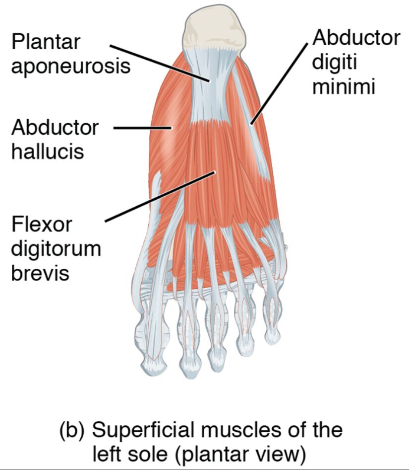
The superficial muscles of the left sole form the outermost layer of the plantar surface. Their plantar view reveals their critical role in foot stability and movement. This section details the labeled structures that define their anatomy and function.
- Abductor hallucis: Positioned along the medial edge, this muscle abducts and flexes the big toe. It supports the medial arch and enhances foot balance.
- Flexor digitorum brevis: Located centrally in the first layer, it flexes the lesser toes. It provides grip strength and aids in weight distribution.
- Abductor digiti minimi: Found along the lateral edge, it abducts and flexes the little toe. It contributes to lateral foot stability and movement.
The superficial muscles of the left sole‘s plantar arrangement ensures robust support. Their labeled anatomy offers a clear insight into their structural and operational roles.
Functional Roles of the Superficial Muscles
The superficial muscles of the left sole are vital for toe flexion and foot support. Their actions in the first plantar layer enhance stability and mobility. This section outlines their specific functional contributions.
- The abductor hallucis abducts and flexes the big toe, supporting the medial arch. It improves balance during weight-bearing activities.
- The flexor digitorum brevis flexes the lesser toes, aiding in gripping the ground. It enhances propulsion and stability during walking.
- The abductor digiti minimi abducts and flexes the little toe, providing lateral support. It assists in maintaining foot alignment on uneven surfaces.
The superficial muscles of the left sole‘s coordinated efforts optimize foot function. Their superficial placement ensures effective support for daily movements.
Clinical Significance and Practical Applications
The superficial muscles of the left sole are often assessed in clinical evaluations of foot health. Their condition directly impacts mobility and stability. This section explores their clinical relevance.
- Weakness in the abductor hallucis can lead to flat feet or hallux valgus. Strengthening exercises help restore arch support and toe alignment.
- Strain in the flexor digitorum brevis may cause toe pain or claw toe deformity. Stretching and conditioning alleviate discomfort and improve function.
- Injury to the abductor digiti minimi can impair little toe movement, affecting lateral stability. Targeted therapy restores flexibility and support.
- Overuse of these muscles may contribute to plantar fasciitis. Rest and rehabilitation prevent further strain and enhance foot health.
- Understanding their anatomy aids in diagnosing conditions like plantar fibromatosis. This knowledge guides effective treatment and preventive strategies.
This insight is valuable for professionals addressing foot concerns. The superficial muscles of the left sole‘s roles underscore the need for precise therapeutic interventions.
Conclusion
The superficial muscles of the left sole, as depicted in the plantar view, highlight the foot’s intricate muscular framework that supports weight-bearing and movement. This article has explored their anatomical structure, diverse functional roles, and clinical significance, providing a thorough understanding of their importance. From the abductor hallucis stabilizing the medial arch to the flexor digitorum brevis enhancing toe grip, each muscle contributes uniquely to foot stability and locomotion. Continued study of these muscles will enhance therapeutic approaches and deepen appreciation for the complex mechanics of the foot.

