The hand is an intricate part of the upper limb, relying on its intrinsic muscles—originating and inserting within the hand—to deliver precise control over the fingers and thumb. This article provides a comprehensive look at the superficial and deep muscles of the left hand, illustrated from both palmar and dorsal perspectives, emphasizing their roles in flexing, extending, abducting, and adducting the distal segments. The detailed images serve as an essential guide for understanding hand anatomy and its functional and clinical importance.
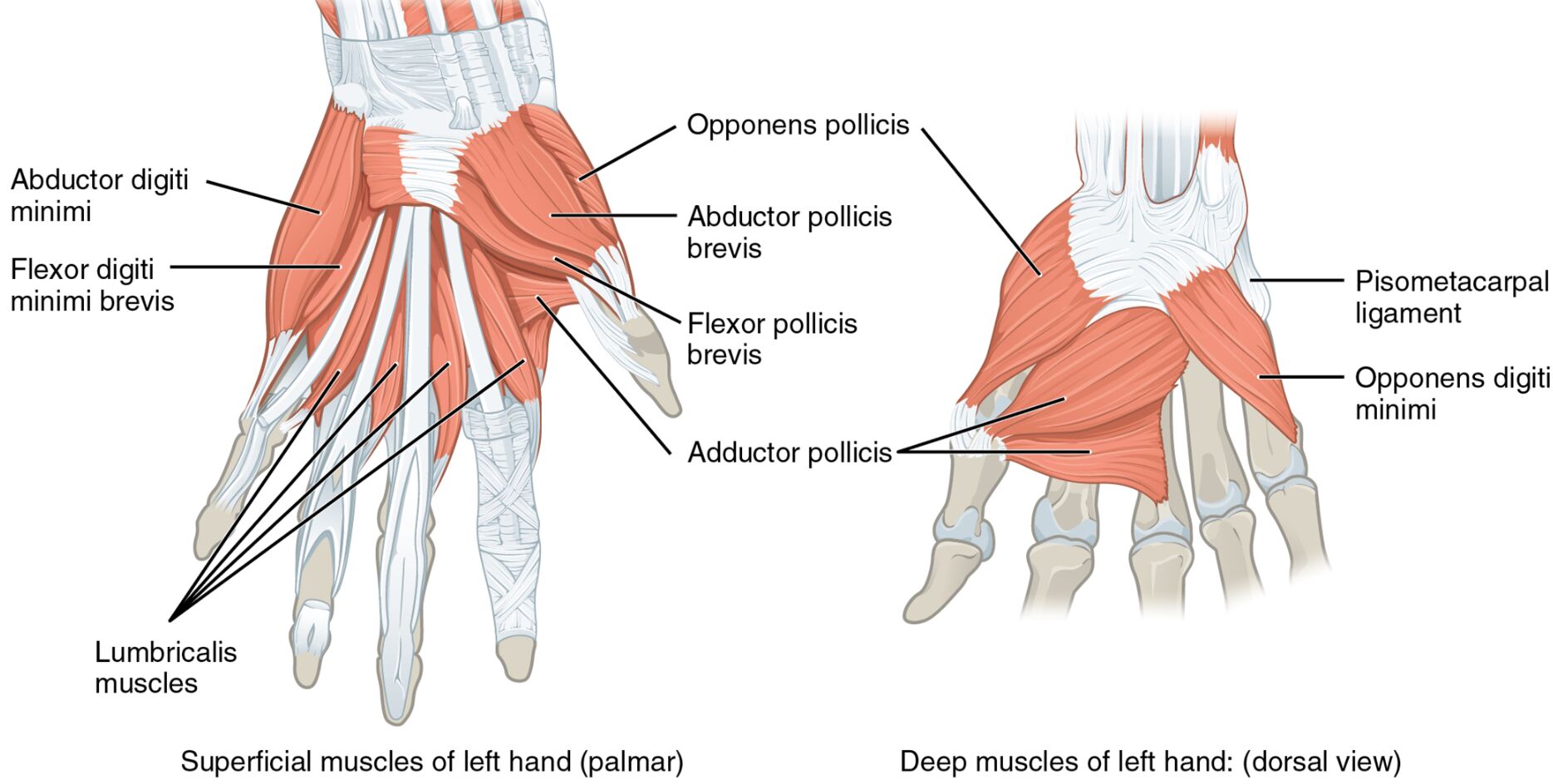
Examining the hand from multiple angles reveals its muscular complexity. The image displays the superficial and deep muscles of the left hand in palmar and dorsal views, with each muscle and related structure clearly labeled.
- Abductor digiti minimi: Originating from the pisiform bone, it abducts the little finger to widen the hand.
- Flexor digiti minimi brevis: Arising from the hamate bone, it flexes the proximal phalanx of the little finger.
- Lumbricalis muscles: Originating from the flexor digitorum profundus tendons, they flex the metacarpophalangeal joints and extend the interphalangeal joints.
- Palmar interossei muscles: Arising from the metacarpal bones, they adduct the fingers toward the middle finger.
- Opponens pollicis: Stemming from the trapezium and flexor retinaculum, it opposes the thumb for grasping.
- Abductor pollicis brevis: Arising from the scaphoid and trapezium, it abducts the thumb away from the hand.
- Flexor pollicis brevis: Originating from the flexor retinaculum and trapezium, it flexes the proximal phalanx of the thumb.
- Adductor pollicis: Stemming from the capitate and metacarpal bones, it adducts the thumb toward the fingers.
- Pisometacarpal ligament: A fibrous band connecting the pisiform to the fifth metacarpal, stabilizing the hypothenar region.
- Opponens digiti minimi: Arising from the hamate bone, it opposes the little finger for enhanced grip.
- Dorsal interossei muscles: Originating from the metacarpal bones, they abduct the fingers away from the middle finger.
- Extensor indicis: Stemming from the ulna, it extends the index finger for individual digit control.
- Extensor pollicis longus: Originating from the ulna, it extends the distal phalanx of the thumb.
- Extensor pollicis brevis: Arising from the radius, it extends and abducts the proximal phalanx of the thumb.
- Abductor pollicis longus: Stemming from the radius and ulna, it abducts and extends the thumb.
Anatomical Overview
Delving into the hand’s muscular structure uncovers a layered complexity. The palmar superficial layer includes the abductor digiti minimi, flexor digiti minimi brevis, lumbricalis muscles, palmar interossei muscles, opponens pollicis, abductor pollicis brevis, flexor pollicis brevis, and adductor pollicis, while the dorsal deep layer features the pisometacarpal ligament, opponens digiti minimi, dorsal interossei muscles, extensor indicis, extensor pollicis longus, extensor pollicis brevis, and abductor pollicis longus.
- The abductor digiti minimi and flexor digiti minimi brevis control little finger movement on the palmar side.
- The lumbricalis and palmar interossei muscles enhance finger flexibility and adduction.
- The opponens pollicis, abductor pollicis brevis, flexor pollicis brevis, and adductor pollicis manage thumb opposition and stability.
- The pisometacarpal ligament and opponens digiti minimi support the hypothenar region.
- The dorsal interossei muscles, extensor indicis, extensor pollicis longus, extensor pollicis brevis, and abductor pollicis longus facilitate finger and thumb extension and abduction.
Functional Roles of Hand Muscles
Understanding the functional dynamics highlights their role in precision. These muscles collaborate to execute fine motor tasks, from thumb opposition to finger abduction, relying on their specific locations.
- The abductor digiti minimi spreads the little finger, aiding hand expansion.
- The flexor digiti minimi brevis flexes the little finger, supporting grip strength.
- The lumbricalis muscles coordinate finger flexion and extension for dexterity.
- The palmar interossei muscles adduct fingers, essential for pinching.
- The opponens pollicis opposes the thumb, crucial for grasping objects.
- The abductor pollicis brevis abducts the thumb, enhancing hand span.
- The flexor pollicis brevis flexes the thumb, vital for holding.
- The adductor pollicis adducts the thumb, reinforcing grip.
- The pisometacarpal ligament stabilizes the hypothenar area during movement.
- The opponens digiti minimi opposes the little finger, improving grip versatility.
- The dorsal interossei muscles abduct fingers, key for hand spreading.
- The extensor indicis extends the index finger, important for pointing.
- The extensor pollicis longus extends the thumb’s distal phalanx, aiding precision.
- The extensor pollicis brevis extends and abducts the thumb’s proximal phalanx.
- The abductor pollicis longus abducts and extends the thumb, supporting grasping.
Clinical Significance
Investigating the clinical implications underscores their practical value. Injuries or dysfunctions in these muscles can disrupt hand function, requiring tailored rehabilitation strategies.
- Strain in the abductor digiti minimi can limit little finger abduction, often treated with therapy.
- The flexor digiti minimi brevis injury may weaken little finger flexion, needing exercises.
- The lumbricalis muscles damage can affect finger coordination, managed with rehabilitation.
- The palmar interossei muscles strain may impair adduction, requiring strengthening.
- The opponens pollicis dysfunction can reduce thumb opposition, treated with physical therapy.
- The abductor pollicis brevis injury may impair thumb abduction, needing care.
- The flexor pollicis brevis strain can weaken thumb flexion, managed with rest.
- The adductor pollicis damage may hinder thumb adduction, necessitating intervention.
- The pisometacarpal ligament injury can destabilize the hypothenar area, treated with support.
- The opponens digiti minimi strain may hinder little finger opposition, requiring therapy.
- The dorsal interossei muscles injury can affect finger abduction, managed with rehabilitation.
- The extensor indicis injury can impair index finger extension, needing care.
- The extensor pollicis longus damage may reduce thumb extension, requiring therapy.
- The extensor pollicis brevis strain can weaken thumb movement, treated with exercises.
- The abductor pollicis longus injury may hinder thumb abduction, necessitating intervention.
Conclusion
The exploration of superficial and deep muscles of the left hand from palmar and dorsal views reveals a sophisticated interplay of anatomy and function. The abductor digiti minimi, flexor digiti minimi brevis, lumbricalis muscles, palmar interossei muscles, opponens pollicis, abductor pollicis brevis, flexor pollicis brevis, adductor pollicis, pisometacarpal ligament, opponens digiti minimi, dorsal interossei muscles, extensor indicis, extensor pollicis longus, extensor pollicis brevis, and abductor pollicis longus each contribute uniquely to fine motor control and hand stability. This dual-view understanding not only enriches knowledge of the hand’s muscular framework but also supports effective management of related injuries, enhancing overall hand health and functionality.

