Streptococcus pyogenes, commonly known as Group A Streptococcus (GAS), is a formidable human pathogen characterized by its unique chain-like arrangement of spherical cells. This Gram-positive bacterium is responsible for a wide clinical spectrum of diseases, ranging from mild pharyngitis to life-threatening invasive infections such as necrotizing fasciitis. Understanding its microscopic morphology and pathogenic mechanisms is essential for rapid diagnosis and effective antibiotic intervention.
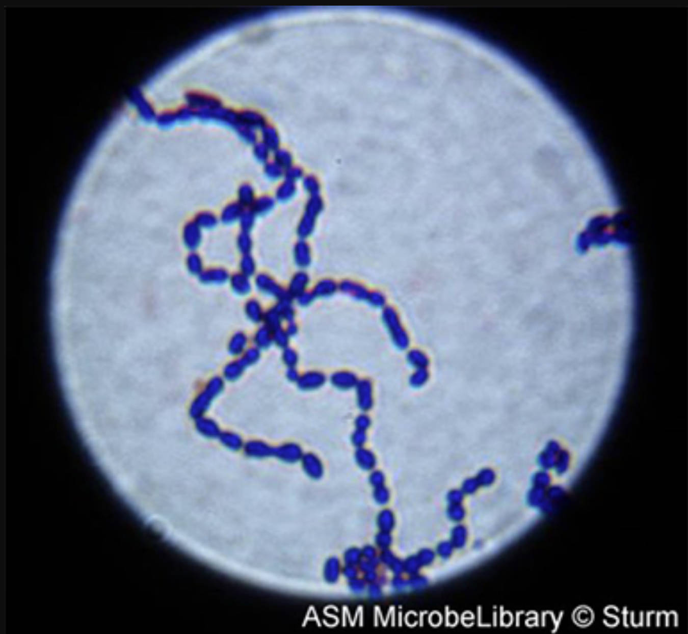
Chains of cocci: This term describes the characteristic arrangement of S. pyogenes where individual spherical bacteria remain attached following cell division along a single plane. Under a light microscope, these formations appear as distinctive, winding strings that allow laboratory professionals to distinguish them from other staphylococcal species that cluster in grape-like groups.
Gram-stained specimen: This diagnostic technique uses crystal violet and safranin stains to categorize bacteria based on the chemical and physical properties of their cell walls. Because S. pyogenes possesses a thick layer of peptidoglycan, it retains the primary purple stain, identifying it as a Gram-positive organism.
Streptococcus pyogenes: Morphology, Pathogenesis, and Clinical Impact of Group A Strep
Streptococcus pyogenes is a ubiquitous bacterium that serves as a primary example of an obligate pathogen that has evolved sophisticated ways to colonize the human body. Classified within the order Lactobacillales, it is a non-motile, non-spore-forming coccus. Its ability to thrive in the human respiratory tract and on the skin surface is facilitated by a complex cell wall structure and an array of secreted virulence factors.
The image provided showcases the “chains of cocci” that define its genus. This morphological trait is more than just a visual marker; it reflects the bacterium’s physiological growth pattern. When GAS divides, the daughter cells do not fully separate, creating long filaments that can help the bacteria adhere more effectively to mucosal surfaces or evade certain host immune responses.
Clinically, this pathogen is most famous for causing “strep throat,” but its reach is far more systemic. If the bacteria breach the initial mucosal barriers, they can enter the bloodstream or deep tissues, leading to severe inflammatory responses. The progression of a GAS infection is often determined by the specific strain’s genetic makeup and the host’s underlying immune status.
Key clinical manifestations associated with Streptococcus pyogenes include:
- Streptococcal pharyngitis (Strep throat)
- Impetigo and cellulitis
- Scarlet fever and Rheumatic fever
- Toxic shock-like syndrome
- Acute glomerulonephritis
The Role of the M Protein and Virulence Factors
The primary virulence factor of Streptococcus pyogenes is the M protein, a hair-like projection extending from the cell wall. This protein is highly anti-phagocytic, meaning it prevents host white blood cells from engulfing and destroying the bacteria. Furthermore, there are over 200 different “M types,” which explains why individuals can suffer from repeated strep infections throughout their lives, as the immune system must learn to recognize each specific variation.
Beyond its physical structure, GAS secretes potent toxins known as streptolysins (O and S). These toxins are responsible for the beta-hemolysis seen on blood agar plates, where the bacteria completely lyse red blood cells, creating a clear halo around the colonies. These enzymes do not just destroy red cells; they also target and kill host immune cells, effectively disarming the local defense mechanisms and allowing the infection to spread rapidly through tissue planes.
Invasive Disease and Post-Streptococcal Complications
In its most aggressive form, S. pyogenes can cause necrotizing fasciitis, a condition colloquially known as “flesh-eating disease.” During this process, the bacteria produce “superantigens” that trigger a massive, non-specific activation of the immune system. This results in a cytokine storm that causes rapid tissue death and systemic organ failure, often requiring emergency surgical debridement and intensive intravenous antibiotic therapy.
Another unique aspect of GAS is the potential for non-suppurative (post-infectious) complications. If a throat or skin infection is not properly treated, the immune system may begin to cross-react with the body’s own tissues due to similarities between the M protein and human proteins. This molecular mimicry can lead to rheumatic heart disease, where the heart valves are permanently damaged, or acute kidney inflammation.
In summary, Streptococcus pyogenes remains one of the most significant bacterial threats to human health due to its versatile morphology and powerful chemical arsenal. From the winding chains of cocci seen in laboratory specimens to the complex immune-evasion strategies of its M protein, this organism requires constant vigilance from healthcare providers. Early identification and adherence to antibiotic protocols remain the gold standard for preventing both the acute symptoms of “strep throat” and the devastating long-term complications of invasive streptococcal disease.

