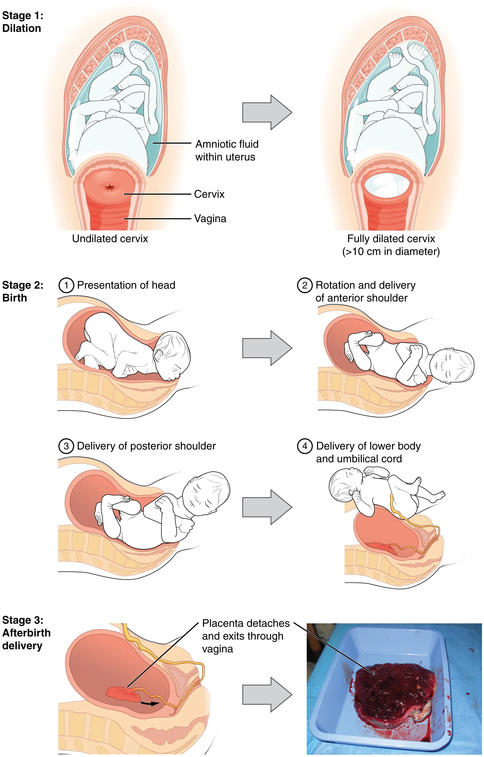
Childbirth is a profound physiological process, meticulously orchestrated into distinct stages to facilitate the safe passage of the newborn from the uterus to the outside world. This comprehensive diagram illustrates the three primary stages of childbirth: Stage 1, focusing on cervical dilation; Stage 2, detailing the birth and expulsion of the newborn; and Stage 3, the delivery of the placenta. Understanding these stages is fundamental for anyone involved in maternal and newborn care, providing a clear framework for monitoring progress and anticipating interventions during labor and delivery.
Key Elements and Stages of Childbirth
Stage 1: Dilation: This initial stage of labor is characterized by the effacement (thinning) and dilation (opening) of the cervix. It is often the longest stage and culminates in the cervix reaching full dilation.
Amniotic fluid within uterus: This fluid surrounds the fetus in the amniotic sac, providing protection and facilitating fetal movement. During labor, the amniotic sac may rupture, releasing this fluid (often referred to as “waters breaking”).
Cervix: The narrow, muscular neck of the uterus that projects into the vagina. Its effacement and dilation are the primary events of the first stage of labor, allowing the baby to pass through.
Undilated cervix: In the initial phase of Stage 1, the cervix is closed or only minimally dilated. It must gradually open to allow the passage of the fetal head.
Vagina: The muscular canal connecting the uterus to the external genitalia. It forms part of the birth canal through which the baby descends during delivery.
Fully dilated cervix (>10 cm in diameter): This marks the end of Stage 1 of labor, indicating that the cervix has opened sufficiently (typically to 10 centimeters) to allow the fetal head to pass into the birth canal.
Stage 2: Birth: This stage begins when the cervix is fully dilated and ends with the complete expulsion of the newborn from the birth canal. It involves active pushing by the birthing person and is typically characterized by distinct phases of fetal descent and rotation.
1 Presentation of head: The initial phase of Stage 2 where the fetal head, usually the largest part, enters and descends through the birth canal. Proper head engagement and flexion are crucial for successful passage.
2 Rotation and delivery of anterior shoulder: After the head is delivered, the baby’s body typically rotates to allow the shoulders to align with the widest part of the pelvic outlet. The anterior shoulder is usually delivered first.
3 Delivery of posterior shoulder: Following the anterior shoulder, the posterior shoulder is delivered. This maneuver often requires careful guidance to prevent injury.
4 Delivery of lower body and umbilical cord: Once the shoulders are clear, the rest of the baby’s body, including the lower limbs, quickly follows. The umbilical cord remains attached at this point, connecting the baby to the placenta.
Stage 3: Afterbirth delivery: This final stage begins immediately after the birth of the baby and concludes with the expulsion of the placenta and fetal membranes. It is crucial for preventing postpartum hemorrhage.
Placenta detaches and exits through vagina: After the baby is born, the uterus continues to contract, causing the placenta to separate from the uterine wall. These contractions then expel the placenta through the vagina.
The Journey of Childbirth
Childbirth is a dynamic and physiological process divided into three main stages. Stage 1, dilation, is often the longest, encompassing the gradual effacement and dilation of the cervix. Uterine contractions, both involuntary and progressively stronger, are the driving force behind these cervical changes. This stage can last for many hours, particularly in first-time birthing persons, and ends when the cervix is fully dilated, typically to 10 centimeters.
Stage 2, birth, commences with full cervical dilation and concludes with the complete delivery of the newborn. This stage involves the descent of the baby through the birth canal, guided by maternal pushing efforts and uterine contractions. The baby undergoes a series of cardinal movements, including engagement, descent, flexion, internal rotation, extension, external rotation, and expulsion, to navigate the pelvis efficiently. The image highlights the crucial steps of head presentation, followed by the delivery of the anterior and posterior shoulders, and finally the rest of the body, with the umbilical cord still attached.
Stage 3, afterbirth delivery, is the shortest but critical stage, beginning after the baby is born and ending with the expulsion of the placenta. After the baby’s delivery, uterine contractions continue, causing the placenta to shear away from the uterine wall. These contractions help to minimize blood loss from the site of placental attachment. Complete expulsion of the placenta and associated membranes is essential to prevent postpartum hemorrhage and infection.
- Each stage of childbirth has specific physiological milestones, and healthcare providers closely monitor these progressions to ensure a safe outcome for both the birthing person and the newborn.
Understanding these sequential stages provides a foundational knowledge of the normal process of labor and delivery.
Conclusion
This comprehensive diagram provides an invaluable visual guide to the three distinct stages of childbirth: dilation, birth, and afterbirth delivery. It clearly illustrates the physiological transformations of the cervix, the intricate movements of the newborn through the birth canal, and the final expulsion of the placenta. A thorough understanding of each stage is paramount for healthcare professionals involved in obstetric care, enabling effective monitoring, support, and intervention to ensure a safe and positive birthing experience.

