The spinal cord serves as a critical conduit for transmitting information between the brain and the rest of the body, relying on a complex network of fiber tracts to facilitate this communication. This diagram illustrates the locations of these spinal fiber tracts and the direction of transmitted information, offering a detailed view of how sensory and motor signals are organized within the spinal cord. Exploring this anatomical layout provides a deeper understanding of how the nervous system coordinates movement, sensation, and reflex actions, making it an invaluable resource for those interested in neurology and physiology.
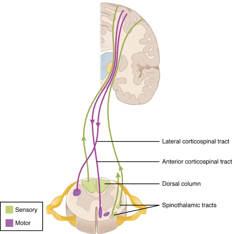
Dorsal column The dorsal column, located in the posterior part of the spinal cord, carries sensory information such as touch, vibration, and proprioception to the brain. It consists of the fasciculus gracilis and fasciculus cuneatus, which relay fine tactile details from the lower and upper body, respectively.
Lateral corticospinal tract The lateral corticospinal tract, found in the lateral region of the spinal cord, transmits motor commands from the brain to control voluntary movements of the limbs. It crosses over to the opposite side of the body at the medullary pyramids, enabling precise muscle coordination.
Anterior corticospinal tract The anterior corticospinal tract, situated in the anterior portion of the spinal cord, also carries motor signals but primarily affects trunk and axial muscles. It remains uncrossed for a portion of its path before some fibers decussate, supporting posture and balance.
Spinothalamic tract The spinothalamic tract, located in the lateral and anterior regions, conveys pain, temperature, and crude touch sensations to the brain. Its fibers cross the midline within a few segments of entry, ensuring rapid relay of critical sensory data.
Spinocerebellar tract The spinocerebellar tract, positioned in the lateral spinal cord, transmits unconscious proprioceptive information to the cerebellum for coordination and balance. It includes the dorsal and ventral divisions, which provide feedback on limb position and movement.
Anatomy of Spinal Fiber Tracts
The spinal cord houses a sophisticated network of tracts for neural communication. This diagram highlights their strategic placement and function.
- The dorsal column occupies the posterior funiculus, ascending sensory fibers to the medulla.
- The lateral corticospinal tract descends from the cortex, crossing at the medulla for contralateral control.
- The anterior corticospinal tract supports axial movements, with partial decussation for bilateral action.
- The spinothalamic tract ascends after crossing, relaying nociceptive and thermal signals.
- The spinocerebellar tract provides real-time feedback, bypassing conscious perception.
Direction of Information Flow
Spinal tracts facilitate bidirectional communication between the brain and periphery. Their directional flow ensures efficient neural signaling.
- Ascending tracts, like the dorsal column, carry sensory input upward to higher brain centers.
- Descending tracts, such as the lateral corticospinal tract, send motor commands downward.
- The spinothalamic tract’s crossing allows contralateral sensory mapping in the thalamus.
- The spinocerebellar tract’s uncrossed path preserves ipsilateral coordination data.
- This organization supports rapid reflexes and voluntary actions alike.
Role of the Dorsal Column in Sensory Processing
The dorsal column is essential for fine sensory discrimination. Its structure supports detailed tactile feedback.
- This tract relays vibration and joint position sense via large myelinated fibers.
- It projects to the nucleus cuneatus and gracilis in the medulla for further processing.
- Damage can lead to loss of proprioception, affecting balance and coordination.
- The tract’s location makes it vulnerable to dorsal column lesions in spinal injuries.
- Rehabilitation focuses on compensatory sensory training in affected cases.
Motor Control via Corticospinal Tracts
The corticospinal tracts govern voluntary movement with precision. This diagram emphasizes their motor dominance.
- The lateral corticospinal tract controls distal muscles, critical for fine motor skills.
- The anterior corticospinal tract stabilizes the trunk, aiding posture during movement.
- Both originate in the primary motor cortex, descending through the internal capsule.
- Upper motor neuron lesions here cause spasticity and weakness on the opposite side.
- Physical therapy targets these tracts to restore motor function post-injury.
Sensory Transmission through the Spinothalamic Tract
The spinothalamic tract handles critical sensory modalities. Its rapid relay enhances survival responses.
- This tract carries pain and temperature signals, crossing within spinal segments.
- It projects to the thalamus, then to the somatosensory cortex for perception.
- Injury can result in contralateral loss of pain sensation, known as a Brown-Séquard syndrome feature.
- The tract’s lateral position makes it susceptible to lateral cord damage.
- Analgesic treatments target this pathway in chronic pain management.
Coordination via the Spinocerebellar Tract
The spinocerebellar tract supports smooth and coordinated movements. Its feedback loop is vital for motor control.
- This tract sends proprioceptive data to the cerebellum without conscious awareness.
- The dorsal division carries information from the lower body, while the ventral covers the upper body.
- Damage disrupts balance and gait, often seen in cerebellar ataxia.
- Its lateral location protects it from some midline injuries.
- Exercises enhancing proprioception aid recovery from tract impairments.
Clinical Relevance of Spinal Tract Mapping
Understanding tract locations aids in diagnosing neurological conditions. This diagram supports clinical applications.
- Lesions in the dorsal column may indicate vitamin B12 deficiency or tabes dorsalis.
- Corticospinal tract damage is assessed via the Babinski sign in upper motor neuron issues.
- Spinothalamic tract injuries are evaluated with pinprick tests for pain loss.
- Spinocerebellar tract dysfunction is tested through heel-to-shin tasks.
- MRI imaging maps these tracts to guide surgical or therapeutic interventions.
Neurotransmitters and Hormonal Influences
Chemical signaling enhances tract function within the spinal cord. These substances modulate neural activity.
- Glutamate excites motor neurons in the corticospinal tracts for movement.
- GABA inhibits excessive signaling, stabilizing sensory input in the spinothalamic tract.
- Serotonin influences pain modulation along ascending pathways.
- Thyroid hormones like T3 and T4 support metabolic health of spinal neurons.
- Imbalances can exacerbate tract-related symptoms, requiring hormonal assessment.
Advances in Spinal Cord Research
Ongoing studies refine our understanding of these tracts. This diagram inspires innovative approaches.
- Diffusion tensor imaging tracks white matter integrity in spinal tracts.
- Stem cell therapy aims to regenerate damaged corticospinal fibers.
- Electrical stimulation enhances motor recovery in tract injuries.
- Gene editing explores repairing congenital tract anomalies.
- These advancements promise improved outcomes for spinal cord patients.
In conclusion, this diagram of spinal fiber tract locations and their direction of transmitted information offers a comprehensive view of the spinal cord’s role in neural communication. From the sensory pathways of the dorsal and spinothalamic tracts to the motor control of the corticospinal tracts and the coordination support of the spinocerebellar tract, each plays a vital part in bodily function. This anatomical insight not only enhances our understanding of the nervous system but also supports clinical diagnostics and emerging treatments, paving the way for better management of spinal cord-related conditions.

