The spinal cord and dorsal root ganglion are integral components of the central and peripheral nervous systems, working together to facilitate sensory and motor functions. This high-magnification micrograph provides a detailed cross-section of the lumbar spinal cord alongside the dorsal root ganglion, revealing the cellular organization and structural complexity of these tissues. Exploring their anatomy offers a deeper appreciation of how they coordinate neural communication and support bodily functions.
Labeled Structures in Spinal Cord and Root Ganglion
This section provides a detailed explanation of each labeled component in the provided image, highlighting their anatomical roles and significance.
Spinal cord The spinal cord is a cylindrical structure extending from the brainstem to the lumbar region, serving as a conduit for motor and sensory signals between the brain and the body. In this cross-section, it reveals gray matter with neuron cell bodies and white matter with myelinated axons, organized into distinct regions like the anterior and posterior horns.
Gray matter The gray matter forms the butterfly-shaped inner region of the spinal cord, containing neuron cell bodies, dendrites, and unmyelinated axons that process sensory and motor information. It is divided into horns—dorsal for sensory input, ventral for motor output, and lateral in some regions—facilitating reflex arcs and signal integration.
White matter The white matter surrounds the gray matter, composed of myelinated axons that form ascending and descending tracts for long-distance neural communication. These tracts, including the corticospinal and spinothalamic tracts, enable coordinated movement and sensory relay to the brain.
Central canal The central canal is a small, fluid-filled channel running longitudinally through the spinal cord’s gray matter, containing cerebrospinal fluid (CSF) that cushions and nourishes the cord. It is a remnant of the neural tube and connects to the ventricular system, aiding in pressure regulation.
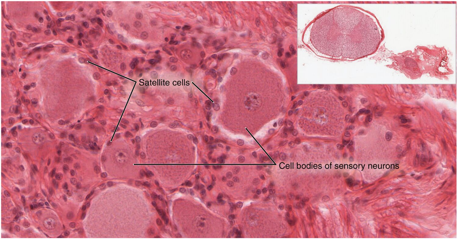
Dorsal root ganglion The dorsal root ganglion is a swelling on the dorsal root of the spinal nerve, housing the cell bodies of unipolar sensory neurons that transmit sensations like touch and pain. This structure is located just outside the spinal cord, with axons extending into the dorsal root to connect with the spinal cord’s sensory pathways.
Dorsal root The dorsal root is the sensory branch of the spinal nerve, carrying axons from the dorsal root ganglion into the spinal cord’s dorsal horn. It contains afferent fibers that relay sensory information from the periphery, playing a key role in reflexes and conscious perception.
Ventral root The ventral root is the motor branch of the spinal nerve, emerging from the spinal cord’s ventral horn and containing axons of motor neurons. These fibers innervate skeletal muscles, facilitating voluntary movement and autonomic functions through efferent signals.
Anatomy of the Spinal Cord and Dorsal Root Ganglion
The spinal cord and dorsal root ganglion exhibit a complex yet organized structure that supports neural function. This anatomical arrangement ensures efficient signal transmission and sensory-motor integration.
- The spinal cord’s gray matter is centrally located, with its horns housing interneurons and motor neurons that process and relay signals.
- White matter tracts are organized into columns—dorsal, lateral, and ventral—each containing specific pathways like the dorsal columns for touch or the lateral corticospinal tract for motor control.
- The central canal, lined with ependymal cells, produces small amounts of CSF locally, contributing to the cord’s buoyancy and protection.
- The dorsal root ganglion, encapsulated by connective tissue, contains sensory neuron cell bodies supported by satellite cells, with axons extending into the dorsal root.
Physiological Role in Neural Communication
The spinal cord and dorsal root ganglion are essential for coordinating sensory input and motor output. Their physiological roles underpin the body’s ability to respond to environmental changes.
- The gray matter processes sensory data from the dorsal root and integrates it with motor commands from the ventral root, enabling reflex actions within milliseconds.
- White matter tracts conduct signals at speeds up to 120 m/s in myelinated fibers, ensuring rapid communication between the brain and spinal levels.
- The central canal’s CSF helps maintain a stable ionic environment, supporting neuronal activity and protecting against mechanical stress.
- The dorsal root ganglion’s unipolar neurons convert peripheral stimuli into electrical impulses, transmitting them via the dorsal root to the spinal cord for further processing.
Clinical Significance and Microscopic Insights
The high-magnification view offers valuable insights into the spinal cord and dorsal root ganglion’s structure and clinical relevance. This level of detail aids in diagnosing and managing neurological conditions.
- Spinal cord injuries affecting the gray or white matter can lead to paralysis or sensory loss, depending on the lesion’s location and extent.
- The dorsal root ganglion is vulnerable to inflammation in conditions like radiculopathy, where nerve root compression causes pain or numbness.
- The 1600x magnification highlights cellular details, such as neuron clustering in the ganglion or myelination in white matter, aiding in the identification of degenerative changes.
- Imaging techniques like MRI or CT myelography assess spinal cord integrity, while nerve conduction studies evaluate dorsal root ganglion function.
Regeneration and Neural Repair
The spinal cord and dorsal root ganglion possess varying capacities for regeneration, influencing recovery after injury. This regenerative potential highlights their adaptability and therapeutic possibilities.
- The spinal cord’s white matter has limited regeneration due to glial scarring, but gray matter neurons can form new synapses under favorable conditions.
- The dorsal root ganglion’s sensory neurons can regenerate peripheral axons, guided by Schwann cells, at a rate of 1-2 mm per day post-injury.
- The central canal’s ependymal cells may contribute to a niche for neural stem cells, potentially aiding repair in experimental therapies.
- Successful regeneration requires minimizing inflammation and scar tissue, which can obstruct axonal regrowth in both structures.
In conclusion, the spinal cord and dorsal root ganglion form a dynamic partnership that supports sensory and motor functions through their intricate anatomy. The detailed insights from this micrograph enhance our understanding of their roles, paving the way for improved diagnostic techniques and innovative treatments for neurological disorders.

