This medical image meticulously illustrates the crucial stages of fertilization, a complex biological event where a sperm cell fuses with an oocyte to initiate the formation of a new organism. Before fertilization can occur, hundreds of capacitated sperm must navigate and penetrate protective layers surrounding the oocyte. This intricate process culminates in the fusion of genetic material, marking the beginning of embryonic development.
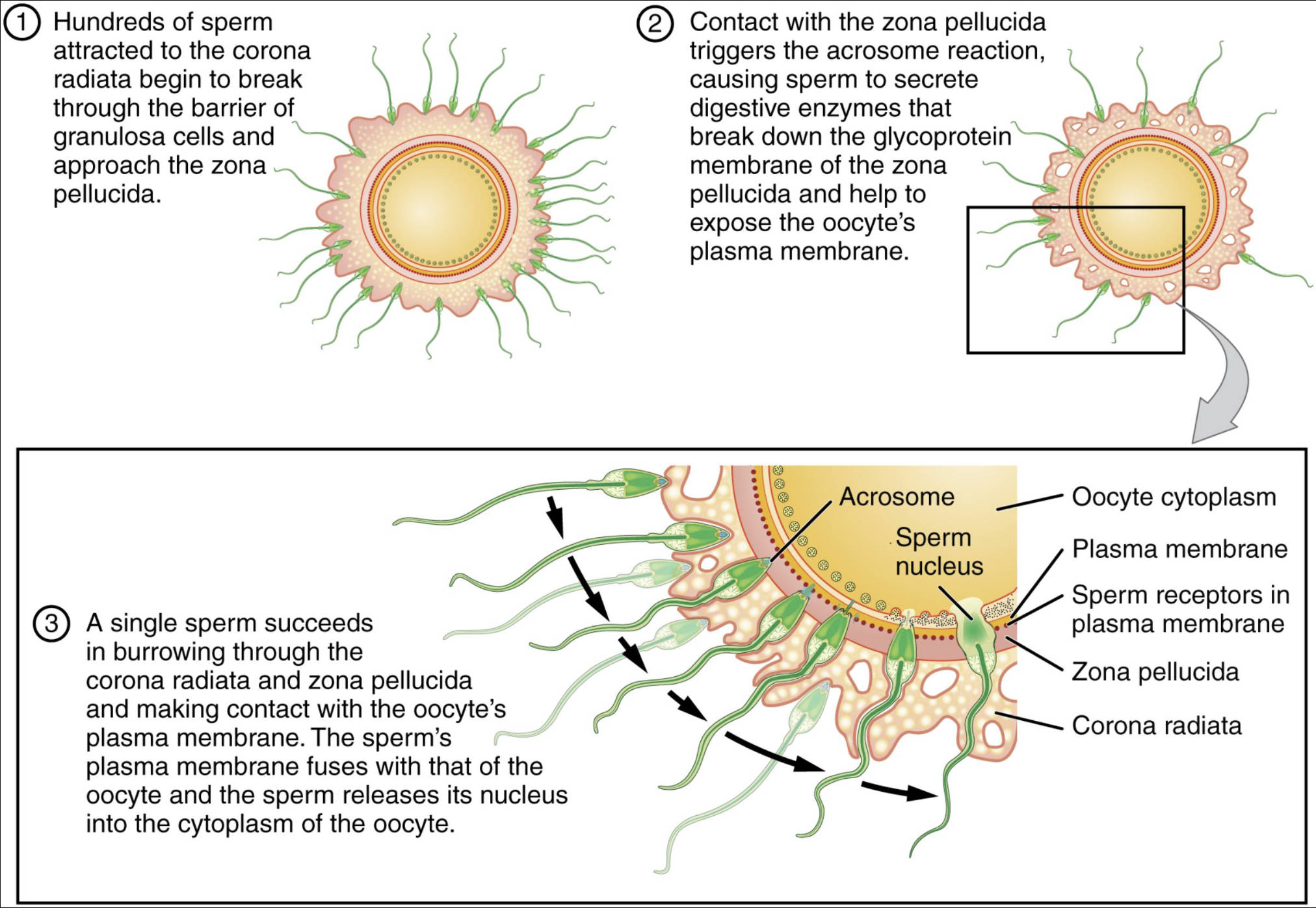
Acrosome: The acrosome is a cap-like organelle located in the anterior part of the sperm head. It contains hydrolytic enzymes, such as hyaluronidase and acrosin, which are essential for breaking down the protective layers of the oocyte, specifically the corona radiata and zona pellucida, allowing the sperm to reach the oocyte’s plasma membrane.
Oocyte cytoplasm: This refers to the jelly-like substance that fills the oocyte, surrounding the nucleus. It contains all the organelles and molecules necessary for the initial stages of embryonic development once fertilization occurs.
Plasma membrane: The plasma membrane is the outer boundary of both the sperm and the oocyte, composed of a phospholipid bilayer. It plays a critical role in mediating the fusion of the two cells during fertilization.
Sperm nucleus: Located within the head of the sperm, the sperm nucleus contains the paternal haploid set of chromosomes. Upon fusion with the oocyte, the genetic material from the sperm nucleus is released into the oocyte’s cytoplasm to combine with the maternal genetic material.
Sperm receptors in plasma membrane: These are specialized protein molecules embedded in the oocyte’s plasma membrane. They are responsible for recognizing and binding to specific molecules on the surface of the sperm, facilitating the attachment and subsequent fusion of the two cells.
Zona pellucida: The zona pellucida is a thick, extracellular glycoprotein layer that surrounds the oocyte, located outside the oocyte’s plasma membrane but inside the corona radiata. It plays a critical role in species-specific sperm binding, induction of the acrosome reaction, and prevention of polyspermy.
Corona radiata: The corona radiata is the outermost layer of the oocyte, composed of several layers of granulosa cells. These cells provide essential nutrients to the oocyte and form a protective barrier that sperm must penetrate before reaching the zona pellucida.
The journey of sperm towards the oocyte is a remarkable feat of cellular navigation and enzymatic action. As depicted in step 1, hundreds of sperm are initially attracted to the corona radiata, beginning their arduous task of breaking through this cellular barrier to approach the zona pellucida. This initial hurdle requires sustained motility and coordinated enzymatic activity from multiple sperm.
Step 2 illustrates a critical event: contact with the zona pellucida triggers the acrosome reaction. During this reaction, the sperm releases a cascade of digestive enzymes that effectively break down the glycoproteins forming the zona pellucida, thereby exposing the oocyte’s plasma membrane. This enzymatic degradation creates a path for one lucky sperm to penetrate this protective layer.
Finally, in step 3, a single sperm successfully burrows through both the corona radiata and zona pellucida, making direct contact with the oocyte’s plasma membrane. This contact initiates a crucial fusion event, where the sperm’s plasma membrane fuses with that of the oocyte. Subsequently, the sperm nucleus, containing the paternal genetic material, is released into the oocyte cytoplasm, marking the completion of fertilization and the commencement of embryonic development. This entire process is tightly regulated to ensure that only one sperm fertilizes the oocyte, a mechanism known as block to polyspermy.
The Journey of Sperm to Fertilization
Fertilization is the culmination of a series of precisely coordinated events that bring together the male and female gametes. This process not only ensures the combination of genetic material from two parents but also triggers the development of a new individual. Understanding the stages involved is crucial for comprehending reproductive biology and the causes of infertility.
Sperm Capacitation and Penetration of the Corona Radiata
Before sperm can fertilize an oocyte, they must undergo capacitation, a maturation process that occurs in the female reproductive tract. This involves biochemical changes that enhance sperm motility and prepare them for the acrosome reaction. Once capacitated, hundreds of sperm surround the oocyte, initiating the penetration of the corona radiata. This layer of granulosa cells is loosely held together by hyaluronic acid, which is partially dispersed by the hyaluronidase enzyme released by the sperm. While many sperm contribute to this dispersion, only a few successfully reach the zona pellucida.
The Acrosome Reaction and Zona Pellucida Penetration
Upon reaching the zona pellucida, a specific glycoprotein layer, sperm bind to species-specific receptors. This binding triggers the acrosome reaction, an exocytosis event where the outer acrosomal membrane fuses with the sperm plasma membrane, releasing a host of enzymes including acrosin. These enzymes create a tunnel through the zona pellucida, allowing the sperm to pass through. Importantly, the zona pellucida also plays a vital role in preventing polyspermy—the fertilization of an oocyte by more than one sperm—by undergoing a “zona reaction” after the first sperm penetrates, rendering it impenetrable to other sperm.
Fusion of Gametes and Genetic Material Transfer
After penetrating the zona pellucida, the sperm makes contact with the oocyte’s plasma membrane. Fusion of the two plasma membranes ensues, allowing the head and tail of the sperm to enter the oocyte cytoplasm. The oocyte then completes its second meiotic division. The sperm nucleus decondenses and swells to form the male pronucleus, while the oocyte nucleus forms the female pronucleus. These two pronuclei then fuse, combining their haploid sets of chromosomes to form a diploid zygote, marking the completion of fertilization and the initiation of embryonic development. This remarkable sequence ensures the genetic integrity and developmental potential of the new organism.

