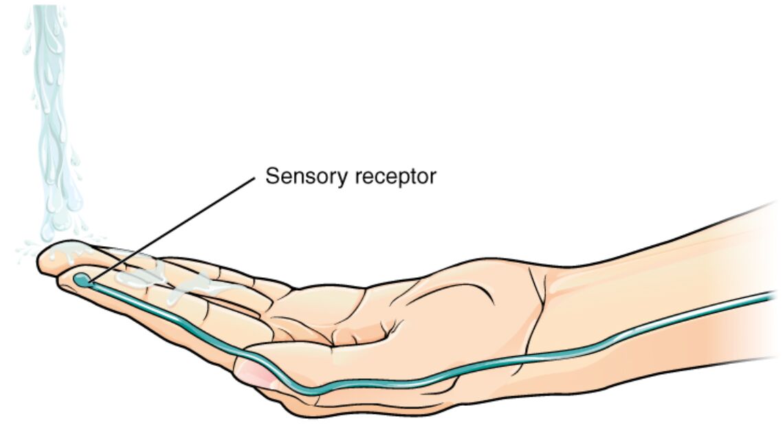
Sensory receptors in the skin serve as the frontline detectors of environmental stimuli, converting physical changes like temperature into neural signals that inform the brain and enable adaptive responses. This diagram depicts a hand exposed to flowing water, emphasizing the activation of cutaneous receptors in the extremity, which are crucial for tactile and thermal perception in everyday interactions. These specialized structures highlight the intricate somatosensory system, where receptors in the epidermis and dermis initiate pathways leading to conscious awareness and reflex actions, essential for protection and fine motor control.
Labeled Parts of the Image
Sensory receptor
The sensory receptor is pointed out at the fingertip, representing embedded structures in the skin that detect stimuli such as temperature, pressure, or pain. These receptors transduce mechanical or thermal energy into electrical signals, initiating graded potentials that may lead to action potentials in afferent neurons.
In-Depth Anatomy of Cutaneous Sensory Receptors
The skin’s sensory apparatus comprises diverse receptor types distributed across layers for multimodal detection. Anatomical variations allow specialization in extremities like hands for heightened sensitivity.
- Thermoreceptors, including cold-sensitive Krause end bulbs and warm-sensitive Ruffini endings, are free nerve endings or encapsulated structures tuned to specific temperature ranges (cold: 5-25°C, warm: 30-45°C).
- Meissner corpuscles in dermal papillae detect light touch and vibration at 50 Hz, with rapid adaptation for dynamic stimuli.
- Pacinian corpuscles, deep in the dermis, sense high-frequency vibrations (250 Hz) via lamellar onion-like capsules that filter low-frequency inputs.
- Merkel cells form complexes with nerve terminals in the basal epidermis, providing sustained responses to pressure for texture discrimination.
- Nociceptors, polymodal free endings, respond to extreme temperatures (>45°C or <5°C), releasing substance P for pain signaling.
Physiological Mechanisms of Sensory Transduction
Receptors convert stimuli into neural codes through ion channel activations. Physiological processes ensure accurate encoding of intensity and location.
- Graded potentials arise from ligand-gated or mechanosensitive channels, such as TRPV1 for heat in warm receptors, depolarizing the membrane proportionally to stimulus strength.
- Action potentials propagate along A-delta (myelinated, fast pain) or C-fibers (unmyelinated, slow pain/itch), with frequency modulation reflecting stimulus intensity.
- Adaptation rates differ: phasic receptors like Meissner quickly cease firing, while tonic like Merkel sustain signals for ongoing stimuli.
- Sensory acuity in extremities benefits from high receptor density (up to 200/cm² in fingertips) and small receptive fields for two-point discrimination.
- Cross-talk with immune cells via cytokines enhances sensitivity during inflammation, amplifying responses to mild stimuli.
Distribution and Specialization in Extremities
Receptors in hands and feet are optimized for interaction with the environment. Specialization supports survival and dexterity.
- Glabrous skin on palms lacks hair but hosts dense Meissner and Merkel receptors for grip and object manipulation.
- Hairy skin on dorsal hands features hair follicle receptors for air movement detection, supplementing thermal cues.
- Depth variation: superficial receptors (epidermis) for fine touch, deeper (hypodermis) for vibration and stretch.
- Innervation by trigeminal (face) or spinal nerves (body) ensures segmented mapping to somatosensory cortex.
- Aging reduces receptor density, contributing to decreased sensitivity, mitigated by neural plasticity.
Neural Pathways from Skin to Brain
Signals from receptors ascend through defined tracts for processing. Pathways integrate peripheral input with central modulation.
- Afferent fibers enter dorsal root ganglia, synapsing in spinal cord lamina for relay via spinothalamic (temperature/pain) or dorsal column (touch/vibration) tracts.
- Thalamic ventral posterolateral nucleus filters signals before cortical projection to postcentral gyrus for perception.
- Descending modulation from periaqueductal gray via opioids can gate pain at spinal levels.
- Cortical somatotopy maps body surfaces disproportionately, with larger areas for sensitive regions like fingers.
- Multisensory integration in association cortices combines thermal with visual/tactile inputs for holistic perception.
Developmental and Evolutionary Aspects
Receptor development stems from ectodermal origins with neural crest contributions. Evolutionary adaptations enhance environmental interaction.
- Embryonic skin innervation occurs by week 8, guided by neurotrophins like NGF for axon growth.
- Postnatal refinement prunes excess synapses, activity-dependent for sharpened receptive fields.
- In mammals, specialized receptors evolved for thermoregulation, differing from reptilian scales lacking such density.
- Genetic factors, like mutations in SCN9A, alter nociceptor function, leading to congenital insensitivity to pain.
- Comparative anatomy shows primates’ enhanced fingertip receptors for tool use and social grooming.
Research Techniques for Studying Skin Receptors
Methods probe receptor function at molecular to systemic levels. Techniques reveal dynamics in health and disease.
- Patch-clamp electrophysiology measures channel currents in isolated receptors, identifying TRPM8 for cold sensation.
- Microneurography records single-fiber activity in vivo, correlating stimuli with neural responses.
- Immunohistochemistry labels proteins like CGRP in nociceptors for localization studies.
- Functional imaging (fMRI) maps cortical activation to specific skin stimuli.
- Biopsies analyze receptor density in conditions like neuropathy using electron microscopy.
Clinical Relevance and Associated Conditions
While the image shows normal function, receptor anomalies underlie disorders. Insights guide diagnostics and treatments.
- Peripheral neuropathies from diabetes damage small fibers, causing thermal hypoesthesia treatable with capsaicin creams.
- Hyperalgesia in fibromyalgia amplifies nociceptor signals, managed by SNRIs like duloxetine.
- Burns disrupt receptors, leading to allodynia where innocuous stimuli evoke pain via central sensitization.
- Congenital disorders like hereditary sensory neuropathies involve Nav1.7 channel mutations, presenting with ulcers from unnoticed injuries.
- Wound healing involves receptor regeneration, promoted by growth factors like EGF.
In summary, the illustrated sensory receptor exemplifies the skin’s vital role as a sensory interface, transducing temperature and other stimuli into actionable neural data for survival and interaction. Exploring these mechanisms deepens appreciation of the somatosensory system and supports advancements in treating sensory disorders, enhancing quality of life through targeted therapies.

