The sensory homunculus is a fascinating representation that illustrates how different parts of the body are mapped onto the brain’s somatosensory cortex, reflecting the sensitivity and density of sensory receptors. This cartoon diagram, positioned adjacent to the cortical region, highlights the disproportionate sizes of body parts based on their sensory innervation, offering a unique insight into neural organization.
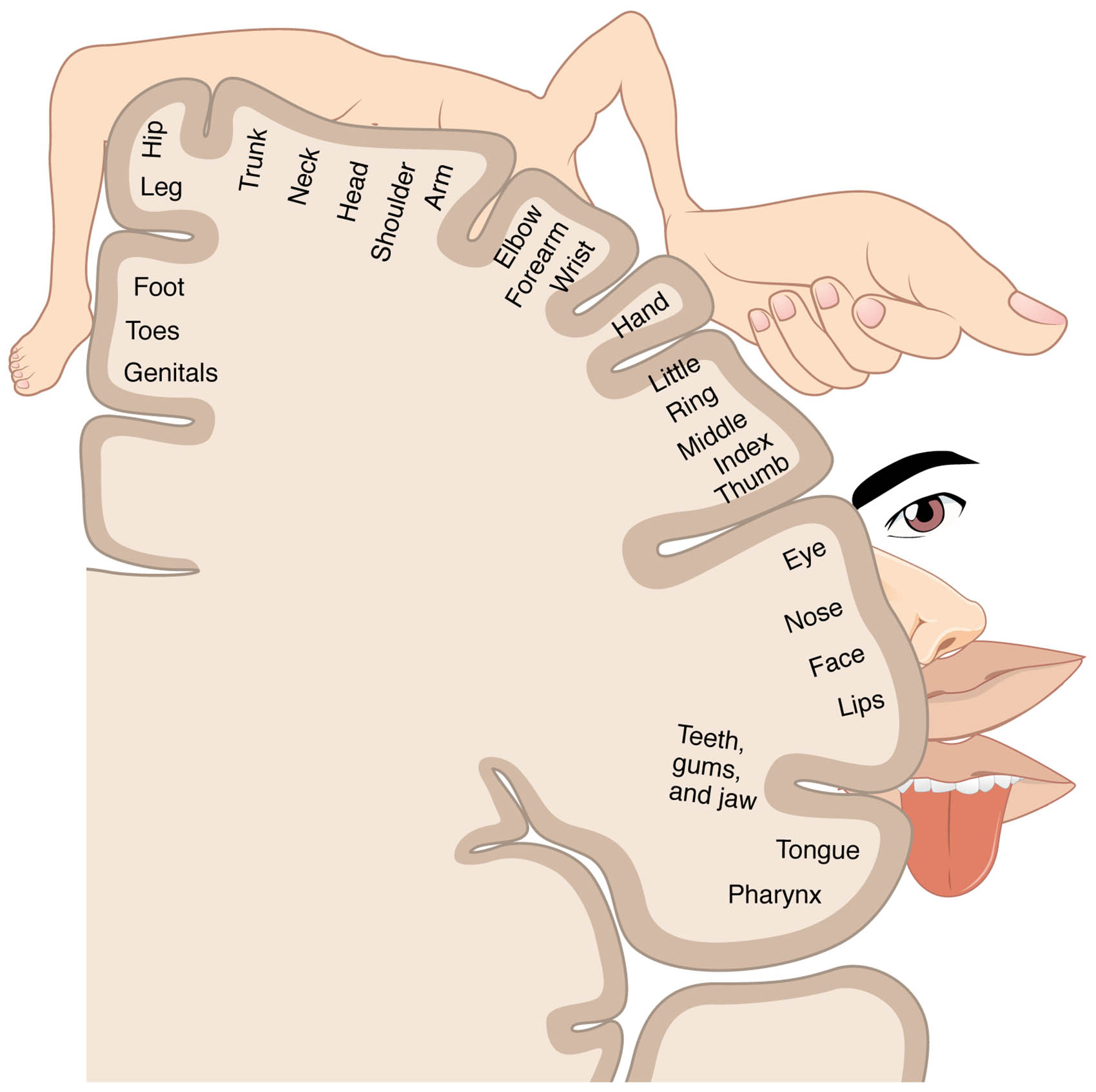
Head and face The head and face are depicted with an exaggeratedly large size on the homunculus, reflecting the high density of sensory receptors in these areas. This region corresponds to the lower part of the postcentral gyrus, processing sensations like touch and pain with great precision.
Hand The hand is shown with an oversized representation, indicating its extensive sensory innervation and fine motor control. It occupies a significant portion of the somatosensory cortex, enabling detailed tactile discrimination.
Arm The arm has a moderately sized depiction, reflecting its sensory input relative to other body parts. Its cortical area processes touch, pressure, and proprioception, with representation tapering toward the shoulder.
Trunk The trunk is represented with a smaller area, corresponding to the lower sensitivity of the torso compared to the extremities. This region of the cortex handles general body sensations like pressure and temperature.
Leg The leg is shown with a moderate size, reflecting its sensory role in movement and balance. Its cortical mapping extends into the parietal lobe, processing sensations from the thigh down to the foot.
Foot The foot has a relatively large representation despite its smaller physical size, due to the importance of sensory feedback for balance and locomotion. This area is located near the top of the somatosensory cortex, adjacent to the leg.
Genitals The genitals are depicted with a small but distinct area, indicating specialized sensory input. This region processes tactile and sexual sensations, mapped near the lower trunk area of the cortex.
Somatosensory cortex The somatosensory cortex, located in the postcentral gyrus of the parietal lobe, is where the sensory homunculus is mapped. It receives and interprets sensory information from the body, organized in a somatotopic manner.
Anatomy of the Sensory Homunculus
The sensory homunculus maps the body’s sensory input onto the somatosensory cortex, creating a distorted yet functional representation. This diagram illustrates how cortical space is allocated based on sensory receptor density.
- The head and face dominate due to the high concentration of receptors in the lips and tongue.
- The hand’s large area supports its role in fine touch and manipulation.
- The arm and leg reflect a gradient of sensitivity, with less cortical space than the hands.
- The trunk covers a broad but less detailed sensory region.
- The foot and genitals highlight specialized sensory zones, despite their smaller physical size.
- The somatosensory cortex is divided into Brodmann areas 3, 1, and 2, each handling different sensory modalities.
- This mapping is contralateral, with the left cortex processing the right body side.
Physiology of Sensory Mapping
The somatosensory cortex processes tactile, proprioceptive, and pain signals, with the homunculus reflecting this activity. This diagram shows how sensory input is organized and interpreted.
- The head and face receive dense innervation, enabling precise facial sensation.
- The hand’s extensive mapping supports detailed touch, like reading Braille.
- The arm and leg process movement-related sensations, aiding coordination.
- The trunk integrates general body awareness, less focused than limb sensations.
- The foot provides balance feedback, critical for posture.
- The genitals handle specialized input, linked to autonomic responses.
- Neural plasticity can adjust these mappings after injury or training.
Role of the Hand in Sensory Precision
The hand’s exaggerated size on the homunculus underscores its role in fine sensory tasks. Its cortical representation supports complex manipulations and tactile feedback.
- The hand’s large area includes separate regions for fingers, thumb, and palm.
- This mapping enhances dexterity, critical for tasks like writing or surgery.
- Receptors like Meissner’s corpuscles detect light touch in this region.
- Damage to the hand’s cortical area can lead to sensory loss or phantom sensations.
- The thumb’s prominence reflects its opposability and tool use.
- Rehabilitation can remap this area after nerve injury.
Role of the Head and Face in Sensory Detail
The head and face’s dominance on the homunculus highlights their sensory richness. This region processes intricate sensations essential for communication and expression.
- The lips and tongue have the highest receptor density, mapped largest on the cortex.
- The head and face handle taste, touch, and facial recognition inputs.
- Trigeminal nerve branches supply this area, enhancing sensitivity.
- Facial paralysis can alter cortical mapping in this region.
- The nose and eyes contribute to multimodal sensory integration.
- This area’s size supports detailed social and environmental interaction.
Clinical Relevance of the Sensory Homunculus
The sensory homunculus provides a framework for diagnosing and treating sensory and motor disorders. This diagram serves as a guide for understanding normal and altered cortical function.
- Stroke affecting the somatosensory cortex can cause numbness in the hand or face.
- Amputation of the arm may lead to remapping or phantom limb sensations.
- Leg or foot sensory loss can result from peripheral neuropathy.
- Trunk desensitization might occur in spinal cord injuries.
- Genitals’ sensory mapping issues can arise in pelvic nerve damage.
- Functional MRI assesses homunculus changes in neurological conditions.
- Physical therapy can stimulate cortical reorganization post-injury.
In conclusion, the sensory homunculus diagram offers a compelling view of how the somatosensory cortex represents the body, with exaggerated areas like the head and face, hand, and foot reflecting their sensory importance. This unique mapping underscores the brain’s adaptability and specialization, providing a valuable tool for exploring neurological health and function.

