Salmonella typhi is a high-impact human pathogen responsible for millions of cases of enteric fever annually. This scanning electron micrograph provides a high-resolution view of the bacterium’s rod-shaped structure, emphasizing its characteristic grouping and surface texture which are essential for its survival in the human host and its ability to trigger systemic illness.
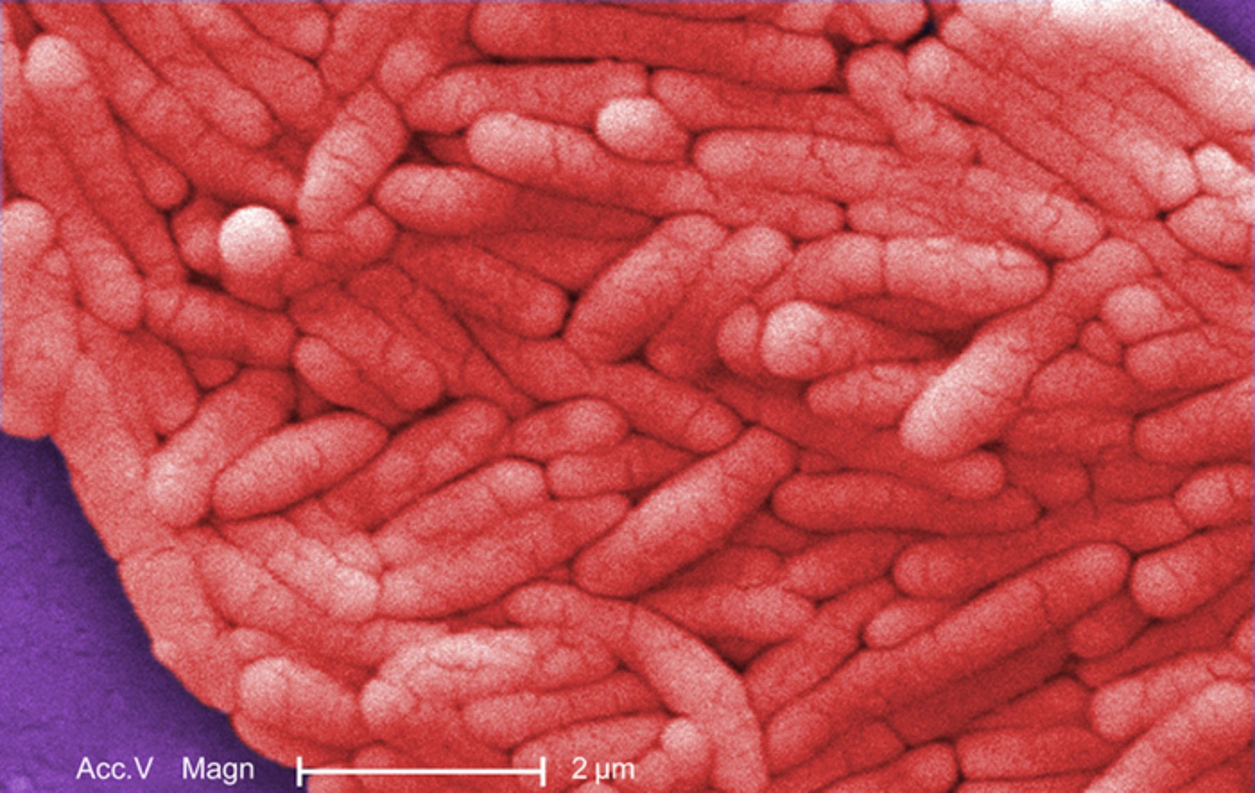
Acc.V Magn: These technical abbreviations stand for Accelerating Voltage and Magnification, representing the specific settings used on the scanning electron microscope. They indicate the intensity of the electron beam and the degree of enlargement required to visualize the intricate surface topography of the bacterial colony.
2 µm: This scale bar provides a crucial reference for the actual physical dimensions of the bacteria displayed in the micrograph. It demonstrates that individual bacilli are microscopic, generally measuring between 0.7 to 1.5 micrometers in width and approximately 2 to 5 micrometers in length.
Salmonella enterica serotype Typhi is a uniquely human-restricted pathogen that distinguishes itself from other salmonellae by its ability to cause severe systemic disease rather than simple localized gastroenteritis. Morphologically, it is a motile, non-spore-forming bacillus that belongs to the Enterobacteriaceae family. It utilizes peritrichous flagella—hair-like appendages distributed over its entire surface—to navigate the complex environment of the human digestive system and eventually infiltrate the bloodstream.
In the laboratory, this Gram-negative organism is identified by its inability to ferment lactose and its production of hydrogen sulfide. The bacteria possess a specialized polysaccharide capsule known as the Vi antigen, which acts as a “virulence” factor by masking other surface antigens from the host’s immune system. This allows the pathogen to bypass initial defenses and establish a foothold in the lymphatic tissues of the small intestine.
The transmission of this pathogen occurs via the fecal-oral route, typically through the consumption of contaminated water or food handled by an asymptomatic carrier. Because humans are the only known reservoir, public health efforts focus heavily on sanitation, water treatment, and the identification of chronic carriers who may unknowingly spread the bacteria.
Essential clinical facts regarding Salmonella typhi include:
- Incubation Period: Symptoms typically emerge 6 to 30 days after initial ingestion.
- The “Step-ladder” Fever: A classic clinical sign where body temperature increases incrementally over several days.
- Systemic Spread: The bacteria primarily colonize the liver, spleen, and bone marrow.
- Chronic Carriage: Approximately 1–5% of recovered patients continue to shed the bacteria in their stool for over a year, often due to colonization of the gallbladder.
Pathophysiology of Typhoid Fever
Once ingested, S. typhi survives the acidic environment of the stomach and reaches the ileum. Here, the bacteria invade the intestinal mucosa through specialized epithelial cells known as M cells. They are subsequently engulfed by macrophages but, rather than being destroyed, they replicate within these immune cells. The bacteria are then transported to the Peyer’s patches, which are organized lymphoid follicles in the gut wall, leading to significant inflammation and potential intestinal perforation in severe cases.
From the lymphatic system, the bacteria enter the thoracic duct and the general circulation, resulting in primary bacteremia. This systemic dissemination allows the pathogen to reach the “RE system” (reticuloendothelial system), specifically the liver and spleen, causing them to enlarge (hepatosplenomegaly). During the second week of infection, a secondary, more intense bacteremia occurs, which is when the classic clinical symptoms of typhoid fever become most pronounced.
Clinical Presentation and Complications
Patients often present with a dull headache, malaise, and a dry cough, followed by high-grade fever. A hallmark physical finding, though present in only about 25% of cases, is the appearance of “rose spots”—faint, salmon-colored macules on the trunk. As the disease progresses, gastrointestinal symptoms may shift from constipation in the early stages to “pea-soup” diarrhea in the later stages.
If left untreated, the inflammatory process in the Peyer’s patches can lead to necrosis, causing life-threatening intestinal hemorrhages or peritonitis. Neurological complications, such as “muttering delirium” or coma vigil, reflect the severe toxemia associated with advanced infection. Fortunately, modern diagnostic tools like blood cultures and the Widal test (though the latter has lower specificity) allow for timely intervention.
Treatment Challenges and Prevention
The management of S. typhi has become increasingly complex due to the emergence of multidrug-resistant (MDR) and extensively drug-resistant (XDR) strains. Historically, chloramphenicol was the treatment of choice, but increasing antibiotic resistance has forced a shift toward fluoroquinolones and third-generation cephalosporins like ceftriaxone. In areas where resistance is high, azithromycin is now frequently utilized as a primary outpatient therapy.
Prevention remains the most effective strategy for controlling the spread of the disease. Two types of vaccines are currently available: the live-attenuated oral vaccine (Ty21a) and the injectable Vi capsular polysaccharide vaccine. More recently, typhoid conjugate vaccines (TCVs) have been introduced, providing longer-lasting immunity and being suitable for younger children. Combined with improvements in global infrastructure and hygiene, these medical advancements aim to eventually eradicate the threat posed by this resilient bacillus.

