The human eye is a dynamic organ, constantly shifting its focus through rapid movements known as saccades to explore and interpret the world around us. This diagram illustrates the saccadic eye movements of an individual studying a face, highlighting the concentrated gaze on key features like the eyes and mouth, with numerous paths indicating active visual scanning. Understanding these patterns offers valuable insights into visual processing and the brain’s role in coordinating eye movements, making this an essential topic for those interested in ocular and neurological health.
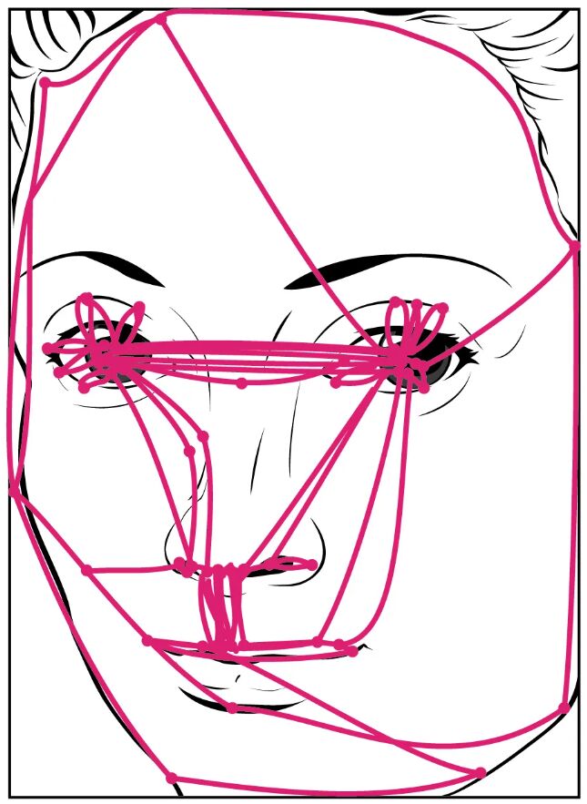
Eyes The eyes are the focal points of gaze in this diagram, attracting significant attention due to their role in facial recognition and emotional expression. Saccades frequently target this area as the brain seeks to gather detailed visual information about the person’s intent or mood.
Mouth The mouth serves as another critical region for saccadic movements, reflecting its importance in speech and emotional cues. The diagram shows repeated shifts to this area, indicating the brain’s effort to analyze lip movements or expressions.
Nose The nose, though less frequently targeted, is part of the saccadic pattern as it provides contextual alignment for facial features. Its inclusion in the gaze path helps the brain map the face’s overall structure during visual exploration.
Face outline The face outline encompasses the entire structure, guiding the overall direction of saccadic movements across the diagram. It represents the boundary within which the eyes navigate to connect major features like the eyes and mouth.
Anatomy of Saccadic Eye Movements
Saccadic movements involve coordinated action within the visual system. This diagram showcases their role in facial analysis.
- Saccadic eye movements are quick, involuntary shifts that reposition the gaze to new points of interest.
- These movements are controlled by the frontal eye fields and superior colliculus in the brain.
- The extraocular muscles, including the lateral rectus and medial rectus, execute these rapid shifts.
- Each saccade lasts about 20-50 milliseconds, too fast for detailed vision during the movement.
- The brain suppresses visual processing during saccades to avoid motion blur.
Neural Control of Saccades
The brain orchestrates saccadic movements with precision. This process ensures efficient visual scanning of complex stimuli.
- The frontal eye fields initiate voluntary saccades, planning the next gaze target.
- The superior colliculus refines these movements, integrating sensory and motor signals.
- The cerebellum adjusts saccade accuracy, correcting for overshoots or undershoots.
- Neural pathways involve the oculomotor, trochlear, and abducens nerves.
- Dopamine and acetylcholine modulate saccade speed and accuracy in these regions.
Role in Facial Recognition
Saccades play a key role in studying faces, as shown in the diagram. Their patterns reveal how we process social cues.
- The concentration on the eyes reflects their importance in detecting gaze direction and emotion.
- The mouth is scanned for speech articulation or subtle expressions like smiles.
- The nose and face outline provide spatial context, aiding in holistic face perception.
- These movements occur 2-3 times per second during active viewing.
- Repeated paths indicate the brain’s prioritization of socially relevant features.
Physiological Benefits of Saccadic Movements
Saccades enhance visual efficiency beyond facial analysis. Their rapid nature supports broader environmental awareness.
- They allow the fovea, the retina’s high-acuity region, to focus on critical details.
- This scanning prevents visual fatigue by distributing attention across the visual field.
- Saccades help track moving objects, like a ball in flight, by predicting trajectories.
- They contribute to spatial memory, aiding navigation in unfamiliar settings.
- Thyroid hormones like T3 and T4 influence overall metabolic support for eye movement.
Clinical Assessment of Saccadic Function
Evaluating saccades is a valuable diagnostic tool. This diagram supports understanding their clinical relevance.
- Smooth pursuit and saccade tests assess neurological health, especially in the brainstem.
- Abnormal saccades, like hypometric jumps, may indicate cerebellar dysfunction.
- Conditions like Parkinson’s disease show delayed or fragmented saccades.
- Eye movement recordings use infrared technology to quantify patterns.
- Results guide treatments, such as physical therapy for coordination issues.
Impact of Fatigue and Stress
Fatigue and stress alter saccadic performance. This diagram prompts exploration of these effects.
- Prolonged visual tasks increase saccade latency, reducing accuracy.
- Stress activates the sympathetic nervous system, potentially speeding up saccades.
- Sleep deprivation disrupts the cerebellum’s fine-tuning of eye movements.
- Chronic stress may lead to irregular gaze patterns, affecting focus.
- Rest and hydration help restore normal saccadic function.
Developmental Aspects of Saccades
Saccadic movements evolve from infancy through adulthood. This diagram reflects mature patterns.
- Newborns exhibit jerky saccades, refining with visual experience by age one.
- Children develop targeted saccades, improving reading and face recognition.
- Adult patterns, as shown, prioritize key facial features efficiently.
- Aging slows saccade velocity, with compensatory strategies emerging.
- Early intervention for delays supports optimal visual development.
Technological Applications
Advances in technology leverage saccadic data. This diagram inspires innovative uses.
- Eye-tracking devices map saccades for usability testing in design.
- Virtual reality systems use saccade patterns to enhance immersive experiences.
- Neurofeedback trains saccade control for rehabilitation post-stroke.
- Research employs saccades to study attention deficits in ADHD.
- These applications expand the diagram’s relevance in modern medicine.
In conclusion, this diagram of saccadic eye movements provides a fascinating glimpse into how the eyes actively explore a face, focusing on key features like the eyes and mouth through rapid shifts. These movements, controlled by intricate neural networks, are essential for visual processing and social interaction, offering a window into brain function and eye health. By studying these patterns, we can better appreciate their role in daily life and leverage this knowledge for clinical assessments and technological advancements, ensuring a deeper understanding of human vision.

