The Q-angle, or quadriceps angle, is a critical measurement in human anatomy that quantifies the lateral deviation of the femur from the vertical line of the tibia, reflecting the alignment of the lower limb. Adult females typically exhibit a larger Q-angle due to their wider pelvis, which influences the biomechanics of the knee and hip joints. This article delves into the anatomical structure of the Q-angle, its physical implications, and its functional and clinical significance in the human body.
Labeled Parts of the Q-Angle: Detailed Explanations
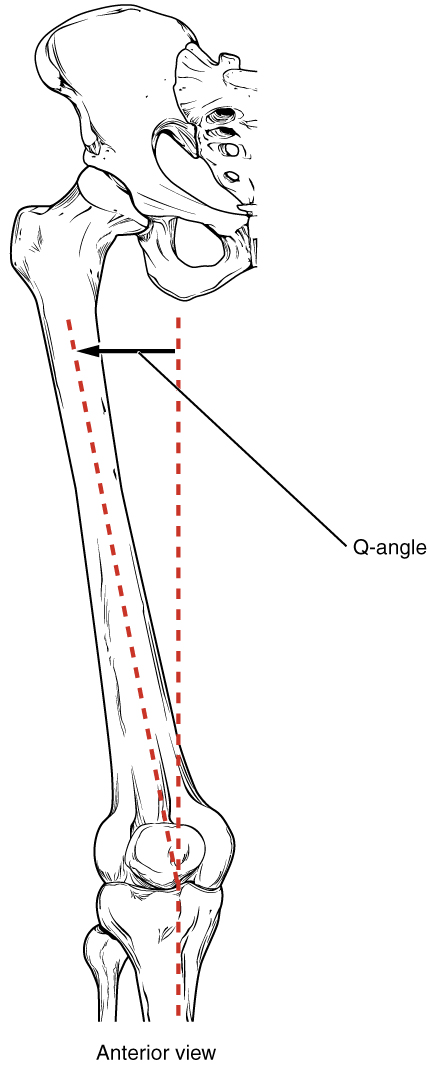
Q-Angle
The Q-angle is the angle formed by two imaginary lines: one drawn from the anterior superior iliac spine (ASIS) to the center of the patella, and another from the center of the patella to the tibial tuberosity. This measurement, typically larger in females (16–18 degrees) than in males (11–14 degrees), reflects the biomechanical alignment of the lower limb and influences knee joint stability.
Anatomical Structure of the Q-Angle
The Q-angle is a key indicator of lower limb alignment, influenced by the skeletal structure of the pelvis, femur, and tibia. This section explores its anatomical basis and the factors contributing to its variation.
- The Q-angle is measured by drawing a line from the ASIS, a bony landmark on the pelvis, to the center of the patella, and a second line from the patella to the tibial tuberosity, a prominence on the proximal tibia.
- A wider pelvis, as seen in females, shifts the femur laterally, increasing the Q-angle and altering the pull of the quadriceps muscle on the patella.
- The femur’s neck-shaft angle, typically 125 degrees, contributes to the Q-angle by determining the femur’s lateral orientation relative to the pelvis.
- The patella, a sesamoid bone within the quadriceps tendon, acts as the pivot point for the Q-angle, influencing the direction of force exerted by the quadriceps on the tibia.
- The tibia’s vertical alignment serves as the reference for the Q-angle, with deviations in tibial torsion or foot positioning potentially affecting the measurement.
Physical Implications of the Q-Angle
The Q-angle’s magnitude has significant physical implications for lower limb biomechanics, particularly in how forces are distributed across the knee joint. This section examines its physical characteristics and their effects on the body.
- A larger Q-angle, common in females due to their wider pelvis, increases the lateral pull on the patella, potentially leading to patellar maltracking or instability.
- In males, a smaller Q-angle results in a more vertical quadriceps pull, which may enhance knee joint stability but can contribute to greater compressive forces on the medial knee compartment.
- The Q-angle affects the alignment of the patellofemoral joint, where the patella articulates with the femur, influencing the distribution of forces during knee flexion and extension.
- Variations in the Q-angle can alter the biomechanics of the hip and ankle joints, as the femur’s lateral deviation impacts the overall alignment of the lower limb.
- The Q-angle’s measurement can be influenced by soft tissue factors, such as quadriceps muscle tightness or ligament laxity, which may exaggerate or reduce the angle in dynamic conditions.
Functional Role of the Q-Angle in the Human Body
The Q-angle plays a crucial role in the biomechanics of the lower limb, affecting movement, stability, and joint health. This section highlights its functional significance in daily activities and locomotion.
- The Q-angle determines the line of pull of the quadriceps muscle, which is essential for knee extension during activities like walking, running, and climbing stairs.
- A larger Q-angle can increase lateral stress on the patella, potentially affecting knee joint stability and contributing to wear on the patellofemoral cartilage over time.
- The Q-angle influences the distribution of forces across the knee joint, with a more vertical alignment in males reducing lateral patellar displacement during movement.
- It impacts the overall alignment of the lower limb, affecting gait efficiency and the coordination between the hip, knee, and ankle joints during locomotion.
- The Q-angle’s role in patellar tracking ensures smooth movement of the patella within the femoral groove, which is critical for maintaining knee joint function during dynamic activities.
Clinical Significance of the Q-Angle
The Q-angle is a valuable measurement in clinical practice, as abnormalities in its magnitude can lead to various musculoskeletal conditions. This section explores its relevance in diagnosing and treating lower limb disorders.
- An increased Q-angle, often seen in females, is associated with conditions like patellofemoral pain syndrome, where excessive lateral pull on the patella causes anterior knee pain.
- A Q-angle greater than 20 degrees can contribute to patellar instability or dislocation, particularly in individuals with hypermobility or weak medial quadriceps muscles.
- In orthopedics, the Q-angle is used to assess lower limb alignment in patients with knee osteoarthritis, as a larger angle may exacerbate medial compartment degeneration.
- Abnormal Q-angles can result from structural variations, such as femoral anteversion or tibial torsion, which may require corrective interventions like physical therapy or surgery.
- Clinicians measure the Q-angle to guide rehabilitation strategies, such as strengthening the vastus medialis oblique muscle to counteract lateral patellar tracking and improve knee stability.
In conclusion, the Q-angle is a fundamental measurement in understanding lower limb biomechanics, reflecting the intricate relationship between the pelvis, femur, and tibia. Its variation between males and females, driven by differences in pelvic width, underscores its importance in both functional and clinical contexts, making it a critical focus for anatomical study and medical practice.

