The pulmonary circuit is a vital component of the circulatory system, responsible for oxygenating blood as it travels through the lungs. This diagram illustrates the pathway from the right ventricle to the pulmonary trunk, pulmonary arteries, capillaries, and back via the pulmonary veins to the left atrium, highlighting the process of gas exchange that sustains life.
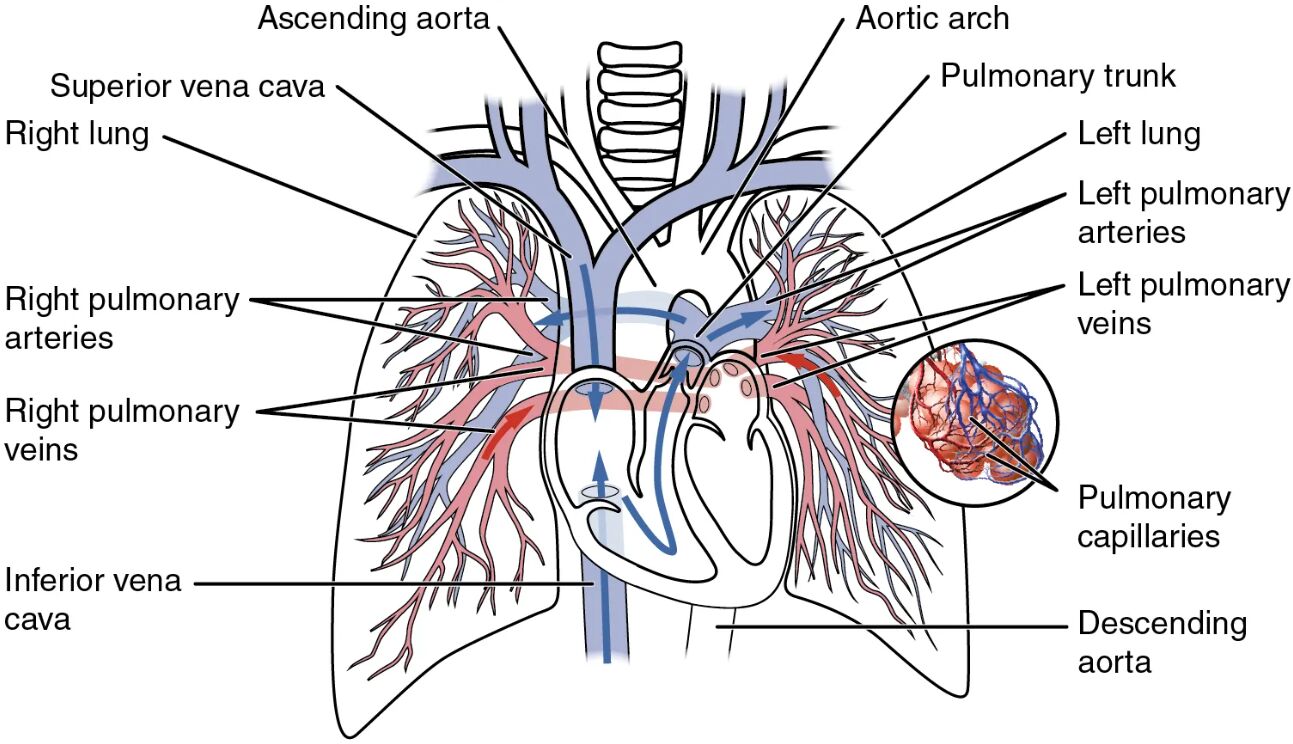
Right ventricle This chamber pumps deoxygenated blood into the pulmonary circuit to be oxygenated. Its thick muscular walls generate the force needed to propel blood toward the lungs.
Pulmonary trunk This large vessel carries blood from the right ventricle toward the lungs. It splits into the pulmonary arteries to distribute blood for gas exchange.
Pulmonary arteries These vessels transport deoxygenated blood from the pulmonary trunk to the lungs. They branch extensively to reach the pulmonary capillaries where oxygenation occurs.
Pulmonary capillaries These tiny vessels surround the alveoli, facilitating the exchange of oxygen and carbon dioxide. Their thin walls allow efficient diffusion between blood and air.
Lung alveoli These small air sacs are the sites of gas exchange within the lungs. They provide a large surface area for oxygen to enter the blood and carbon dioxide to be expelled.
Pulmonary veins These vessels return oxygenated blood from the lungs to the left atrium. They are unique among veins as they carry oxygen-rich blood.
Left atrium This chamber receives oxygenated blood from the pulmonary veins. It then pumps the blood into the left ventricle to be distributed throughout the body.
Anatomy of the Pulmonary Circuit
The pulmonary circuit begins with the right ventricle, setting the stage for oxygen replenishment. Its design ensures efficient blood flow through the lungs for gas exchange.
- The right ventricle’s muscular structure supports the initial push into the pulmonary trunk.
- The pulmonary trunk acts as a conduit, dividing into the pulmonary arteries.
- Pulmonary arteries carry deoxygenated blood, contrasting with the systemic arteries.
- Capillaries maximize surface area for oxygen diffusion into the bloodstream.
- The circuit’s structure minimizes resistance to maintain steady flow.
Gas Exchange in the Lungs
Pulmonary capillaries and lung alveoli are central to the gas exchange process. Their close proximity enables critical respiratory functions.
- Oxygen from the alveoli diffuses into the capillaries to bind with hemoglobin.
- Carbon dioxide, a waste product, moves from the blood into the alveoli to be exhaled.
- The thin capillary walls enhance the speed of diffusion.
- Alveolar surfactant reduces surface tension, keeping alveoli open.
- This exchange is vital for maintaining adequate oxygen levels in blood.
Pathway of Blood Through the Circuit
The journey from the pulmonary trunk to the pulmonary veins outlines the circuit’s efficiency. Each segment plays a specific role in oxygenation.
- The pulmonary trunk bifurcates to ensure even blood distribution to both lungs.
- Pulmonary arteries branch into smaller vessels, reaching all lung regions.
- Blood flows through capillaries, where gas exchange transforms its composition.
- Pulmonary veins collect oxygenated blood, preparing it for systemic circulation.
- The left atrium receives this blood, completing the pulmonary phase.
Role of the Heart in the Pulmonary Circuit
The right ventricle and left atrium are key cardiac components of the pulmonary circuit. Their coordinated action drives the circulation process.
- The right ventricle pumps with sufficient force to overcome pulmonary resistance.
- Its output matches the body’s oxygen demand during rest or activity.
- The left atrium acts as a reservoir, ensuring smooth flow into the left ventricle.
- Valves between these chambers prevent backflow, maintaining directionality.
- This cardiac support is essential for effective gas exchange.
Clinical Significance of the Pulmonary Circuit
Understanding the pulmonary circuit aids in diagnosing and managing respiratory issues. Its anatomy provides insights into potential health challenges.
- Blockages in pulmonary arteries can lead to pulmonary embolism.
- Capillary damage may impair gas exchange, causing hypoxia.
- Blood oxygenation levels are monitored to assess lung function.
- Alveolar diseases like pneumonia affect the circuit’s efficiency.
- Surgical interventions often target this circuit in congenital heart defects.
The pulmonary circuit exemplifies the body’s remarkable ability to oxygenate blood and remove carbon dioxide, sustaining vital functions. From the right ventricle’s initial push to the left atrium’s reception, this system ensures every tissue receives the oxygen needed, offering a foundation for exploring respiratory and cardiovascular health.

