The posterior view of the sacrum and coccyx offers a detailed look at the lower spine’s foundational elements, crucial for understanding human anatomy. This region plays a vital role in supporting the body’s weight and connecting the spine to the pelvis, making it an essential area of study for grasping skeletal structure and function.
Labels Introduction
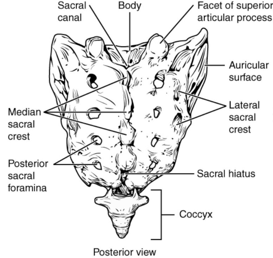
Sacral canal: The sacral canal is a hollow, elongated space running through the center of the sacrum, serving as a passageway for the spinal cord’s continuation and nerve roots. It extends from the sacral hiatus to the upper part of the sacrum, providing protection and support to the lower spinal nerves.
Body: The body of the sacrum forms the central, broad portion of each sacral vertebra, fused together to create a sturdy triangular bone. This structure bears much of the body’s weight and articulates with the lumbar spine above.
Facet of superior articular process: The facet of the superior articular process is a smooth, curved surface on the upper part of the sacrum that allows for articulation with the fifth lumbar vertebra. This joint facilitates limited movement and stability between the spine and sacrum.
Auricular surface: The auricular surface is a rough, ear-shaped area on the lateral side of the sacrum that articulates with the ilium of the pelvis, forming the sacroiliac joint. This joint is critical for transferring weight from the spine to the lower limbs and is stabilized by strong ligaments.
Lateral sacral crest: The lateral sacral crest consists of fused transverse processes on either side of the sacrum, providing attachment points for muscles and ligaments that support pelvic stability. These crests also help in aligning the sacrum with the pelvis.
Median sacral crest: The median sacral crest is a midline ridge formed by the fused spinous processes of the sacral vertebrae, serving as an attachment site for ligaments and muscles. It runs vertically along the posterior sacrum, contributing to its structural integrity.
Posterior sacral foramina: The posterior sacral foramina are a series of openings on the back of the sacrum that allow the passage of the sacral spinal nerves and blood vessels. These foramina are symmetrically arranged and play a key role in the nervous system’s lower spinal communication.
Sacral hiatus: The sacral hiatus is an opening at the lower end of the sacral canal, marking the termination of the canal and providing access for procedures like caudal epidural anesthesia. It is formed by the failure of the lamina of the fifth sacral vertebra to fuse.
Coccyx: The coccyx, or tailbone, is a small, triangular bone at the base of the sacrum formed by the fusion of four coccygeal vertebrae. It serves as an attachment point for ligaments and muscles of the pelvic floor, supporting sitting posture and movement.
Detailed Anatomical Overview
Understanding the Sacrum and Coccyx Structure
The sacrum and coccyx form the foundation of the pelvic girdle, playing a pivotal role in human locomotion and stability. This region supports the weight of the upper body and distributes it to the lower limbs through the sacroiliac joints. Its unique triangular shape and fused vertebrae provide the strength needed to withstand significant mechanical stress.
- The sacral canal houses the dural sac and cauda equina, extending nerve roots that innervate the lower body.
- The body of the sacrum is robust, with a concave anterior surface that enhances its weight-bearing capacity.
- The facet of superior articular process ensures a smooth transition of movement from the lumbar spine, reducing wear on adjacent structures.
Functional Roles in the Body
The sacroiliac joint, formed by the auricular surface, is a synovial joint reinforced by ligaments, allowing minimal movement while maintaining stability. This joint is crucial during activities like walking or lifting, where force transmission is essential. The lateral sacral crest and median sacral crest anchor muscles such as the erector spinae, aiding in posture and spinal alignment.
- The posterior sacral foramina facilitate the exit of sacral nerves, which control bladder, bowel, and lower limb functions.
- The sacral hiatus is a clinically significant landmark, often used for administering anesthesia in surgical procedures.
- The coccyx supports the pelvic floor muscles, including the levator ani, which are vital for maintaining continence and aiding childbirth.
Clinical Relevance and Anatomical Considerations
The sacrum’s structure is integral to understanding conditions like sacral fractures or coccydynia (tailbone pain). The sacral canal can be a site for nerve compression if abnormalities occur, potentially leading to pain or numbness. Proper knowledge of these structures aids in diagnosing and treating lower back issues effectively.
- The body can be assessed for deformities using imaging techniques like X-rays or CT scans.
- The facet of superior articular process may become arthritic, causing localized pain that radiates to the hips.
- The auricular surface’s alignment is critical in cases of sacroiliac joint dysfunction, a common source of lower back pain.
Supporting Structures and Their Importance
The lateral sacral crest and median sacral crest provide attachment for the sacrotuberous and sacrospinous ligaments, which stabilize the pelvis. These crests also serve as landmarks during surgical interventions. The posterior sacral foramina are key in nerve block procedures, offering relief from chronic pain.
- The sacral hiatus is a gateway for caudal epidurals, delivering medication to the epidural space.
- The coccyx can be prone to injury from falls, leading to persistent discomfort that requires medical attention.
The sacrum and coccyx, though small in comparison to other skeletal components, are indispensable for overall body mechanics. Their intricate design supports weight, facilitates movement, and protects vital neural structures. Understanding their anatomy enhances clinical practice and patient care, ensuring accurate diagnosis and effective treatment plans.
Conclusion
The posterior view of the sacrum and coccyx reveals a complex interplay of bones and joints that underpin human posture and mobility. This anatomical region’s study is essential for grasping its role in supporting the body and facilitating nerve function. By exploring its labeled parts and clinical significance, one can appreciate the sacrum and coccyx’s critical contribution to overall health and well-being.

