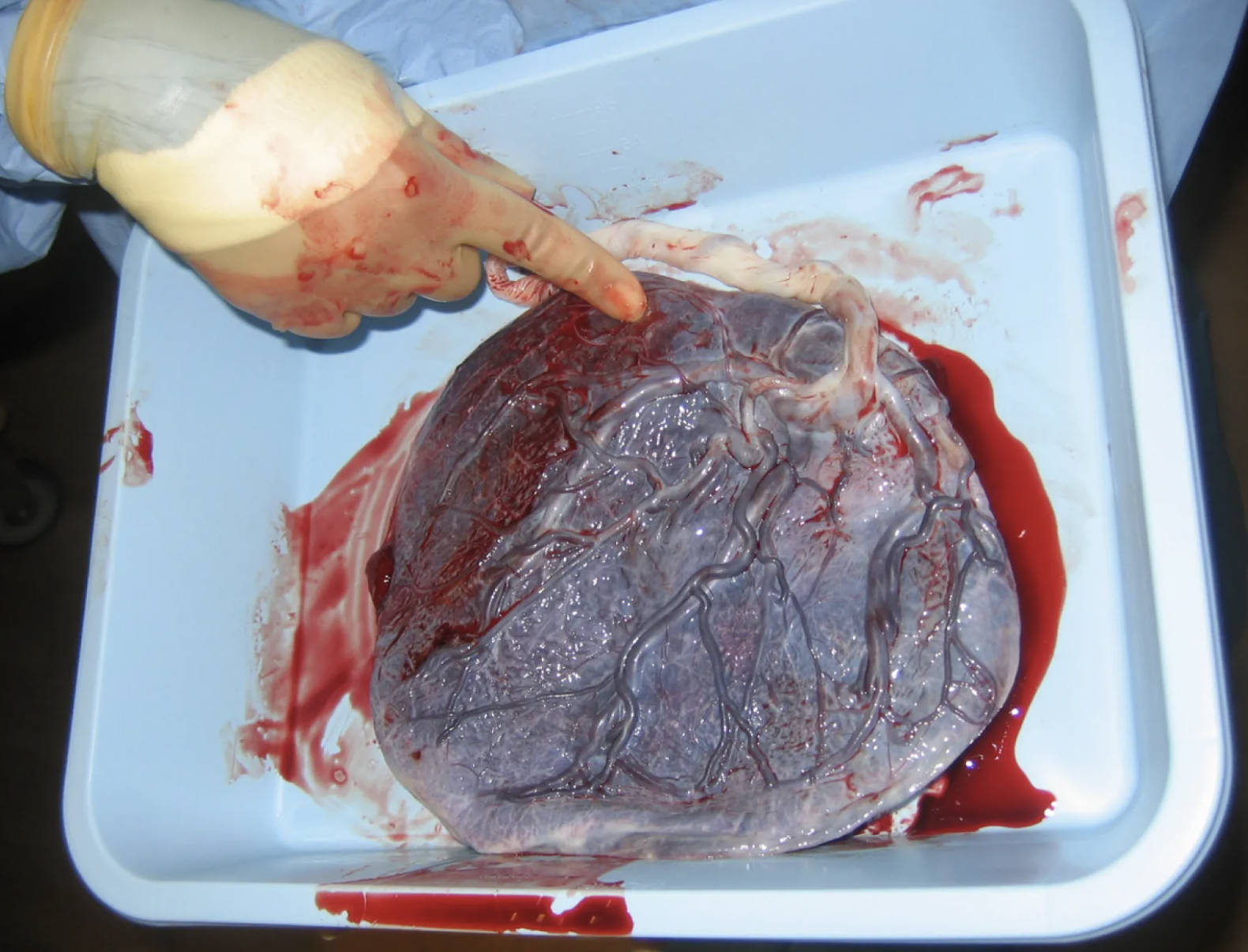
Following childbirth, the placenta, often referred to as the afterbirth, is expelled from the uterus. This image captures a post-expulsion placenta and its attached umbilical cord, viewed from the fetal side. This perspective offers a clear view of the amnion and the intricate network of blood vessels that once connected the fetus to its mother, facilitating vital exchanges throughout pregnancy. Examining the placenta post-delivery is a routine practice in obstetrics, as it can provide crucial insights into the health of both the mother and the newborn, revealing any potential complications that may have occurred during gestation.
Anatomical Features of the Post-Expulsion Placenta
The image shows a post-expulsion placenta and its umbilical cord. While there are no specific labels on this particular image, we can identify and discuss the key anatomical features visible from the fetal side.
Placenta (Fetal Side): This is the large, disc-shaped organ visible in the image. The fetal side is typically smooth and shiny, covered by the amnion. It is characterized by the radiating blood vessels originating from the umbilical cord insertion point. This surface represents the interface where the fetal blood vessels came into close proximity with maternal blood, facilitating nutrient, gas, and waste exchange.
Umbilical Cord: The umbilical cord is the rope-like structure attached centrally (or sometimes eccentrically) to the fetal surface of the placenta. In the image, it is distinctly visible as a white, twisted structure. It typically contains two umbilical arteries and one umbilical vein, encased in Wharton’s jelly, which protects these vessels. After birth, the cord is clamped and cut, leaving a stump on the newborn.
Amnion: The thin, translucent membrane that covers the fetal surface of the placenta and the umbilical cord. In a healthy placenta, the amnion is intact and gives the surface a glistening appearance, reflecting the healthy environment it provided for the developing fetus.
Chorionic Plate: While not explicitly labeled, the chorionic plate is the base of the chorion from which the chorionic villi project into the intervillous space. On the fetal side, this is the area where the umbilical cord vessels branch out, forming the vascular tree visible beneath the amnion.
Blood Vessels (Fetal): The prominent network of vessels spreading across the fetal surface of the placenta originates from the umbilical cord. These are the umbilical arteries and vein, which branch repeatedly to supply the chorionic villi. Their distribution and integrity are crucial for efficient fetal circulation and placental function.
Significance of Placental Examination
The post-expulsion placenta serves as a valuable record of the pregnancy. Its examination can reveal information about fetal growth, potential infections, and circulatory issues. For instance, the color, consistency, and presence of any lesions or infarcts on the placental tissue can indicate underlying conditions. An abnormally large or small placenta, or the presence of calcifications, can be correlated with fetal growth restriction or other developmental concerns.
The insertion point of the umbilical cord is also significant; a central insertion is ideal, while marginal or velamentous insertions can sometimes be associated with complications like vasa previa. The length and twist of the umbilical cord are also noted, as extreme variations can sometimes impact fetal well-being. Furthermore, the presence of accessory lobes (succenturiate lobes) or remnants of membranes can increase the risk of postpartum hemorrhage if not completely removed from the uterus.
- A thorough examination includes assessing the weight, dimensions, and completeness of the placental tissue to ensure no parts are retained within the uterus.
This visual inspection, coupled with histological examination if warranted, offers healthcare providers a comprehensive understanding of the gestational period, aiding in the postnatal care of both mother and infant.
Conclusion
The post-expulsion placenta, as depicted from its fetal side, is more than just an afterbirth; it is a critical organ that supported life and growth throughout pregnancy. Its intricate vascular network and protective membranes are testament to its vital role in maternal-fetal exchange. The routine examination of this expelled organ provides crucial diagnostic information, allowing healthcare professionals to assess the health of the pregnancy and identify any anomalies that may impact the well-being of the newborn and the mother.

