Platelets are essential blood components responsible for clotting and wound healing, originating from specialized cells known as megakaryocytes within the bone marrow. This diagram illustrates the process of platelet formation, detailing the transformation and release of these cell fragments into the bloodstream to maintain hemostasis. Exploring this lifecycle offers valuable insights into the body’s ability to prevent excessive bleeding and support tissue repair.
Key Components of Platelet Formation
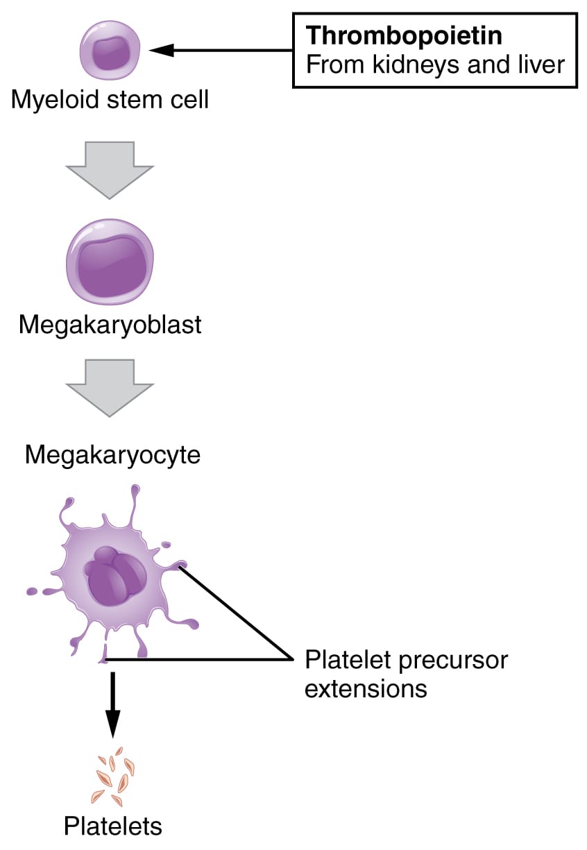
The diagram outlines the stages and structures involved in platelet development.
Megakaryoblast:
The megakaryoblast is the earliest precursor cell in platelet formation, originating from hematopoietic stem cells in the bone marrow. It undergoes rapid division and growth, setting the stage for further differentiation into mature platelets.
Promegakaryocyte:
The promegakaryocyte represents an intermediate stage with increased cytoplasm and the beginning of polyploidy, where the cell prepares for platelet production. It continues to develop within the bone marrow, accumulating organelles needed for future platelet function.
Megakaryocyte:
The megakaryocyte is a large, multinucleated cell that produces platelets through cytoplasmic fragmentation, residing in the bone marrow. It extends cytoplasmic processes called proplatelets into sinusoids, releasing thousands of platelets into circulation.
Proplatelets:
Proplatelets are elongated cytoplasmic extensions from megakaryocytes that serve as the direct precursors to platelets, protruding into blood vessels. They undergo fission to release individual platelets, ensuring a steady supply for hemostatic needs.
Platelets:
Platelets are small, anucleate cell fragments released from proplatelets, circulating for about 7-10 days with a count of 150,000-450,000 per microliter. They play a critical role in clot formation by aggregating at injury sites and releasing clotting factors.
Bone marrow sinusoids:
Bone marrow sinusoids are specialized vascular channels where megakaryocytes release proplatelets, facilitating platelet entry into the bloodstream. These porous structures allow direct access to circulation, supporting rapid platelet distribution.
The Anatomical and Physiological Role of Platelet Formation
Platelet formation begins with the megakaryoblast, a committed progenitor in the bone marrow, stimulated by thrombopoietin from the liver and kidneys in response to low platelet counts. The promegakaryocyte stage marks the onset of polyploidy, where DNA replication occurs without cell division, preparing the cell to become a megakaryocyte. This large cell, with its multiple nuclei, extends proplatelets into bone marrow sinusoids, where they fragment into functional platelets.
These platelets are crucial for hemostasis, adhering to damaged endothelium via glycoproteins like GPIb and GPIIb/IIIa, triggered by exposure to collagen. Hormones such as T3 and T4 from the thyroid gland indirectly influence metabolic demands, affecting overall hematopoiesis. The process ensures a constant platelet supply, with old platelets removed by the spleen, maintaining a balance critical for clotting and tissue repair.
- Production Regulation: Thrombopoietin binds to c-Mpl receptors on megakaryocytes; levels rise after bleeding or platelet destruction.
- Release Mechanism: Proplatelets shear off in response to blood flow; sinusoids enhance platelet maturation.
- Functional Role: Platelets release ADP and serotonin; granules store von Willebrand factor for adhesion.
Physical Characteristics and Clinical Relevance
The physical evolution of platelets reflects their specialized role, observable through microscopic analysis. Megakaryoblasts are small and round, while megakaryocytes grow large with lobulated nuclei, and proplatelets appear as thread-like extensions. Mature platelets are disc-shaped with purple-staining granules, designed for rapid activation.
Clinically, this diagram aids in diagnosing platelet disorders. Low platelet counts, or thrombocytopenia, may result from bone marrow failure or increased destruction, as in immune thrombocytopenic purpura (ITP), requiring corticosteroids or IVIG. Elevated counts, seen in essential thrombocythemia, increase clotting risk, managed with aspirin or cytoreductive therapy. Complete blood counts (CBC) and bone marrow biopsies assess production, guiding interventions.
- Diagnostic Tools: Mean platelet volume (MPV) indicates platelet size; flow cytometry detects immature platelet fractions.
- Therapeutic Approaches: Platelet transfusions address severe bleeding; eltrombopag stimulates megakaryopoiesis in aplastic anemia.
Conclusion
The platelet formation diagram reveals the intricate process by which megakaryocytes generate platelets, ensuring the body’s ability to clot blood and heal wounds effectively. From the bone marrow’s nurturing environment to the release into circulation, each stage underscores the precision of hematopoiesis. Understanding this cycle enhances the ability to diagnose and treat platelet-related conditions, highlighting the critical role these tiny fragments play in maintaining vascular integrity and overall health.

