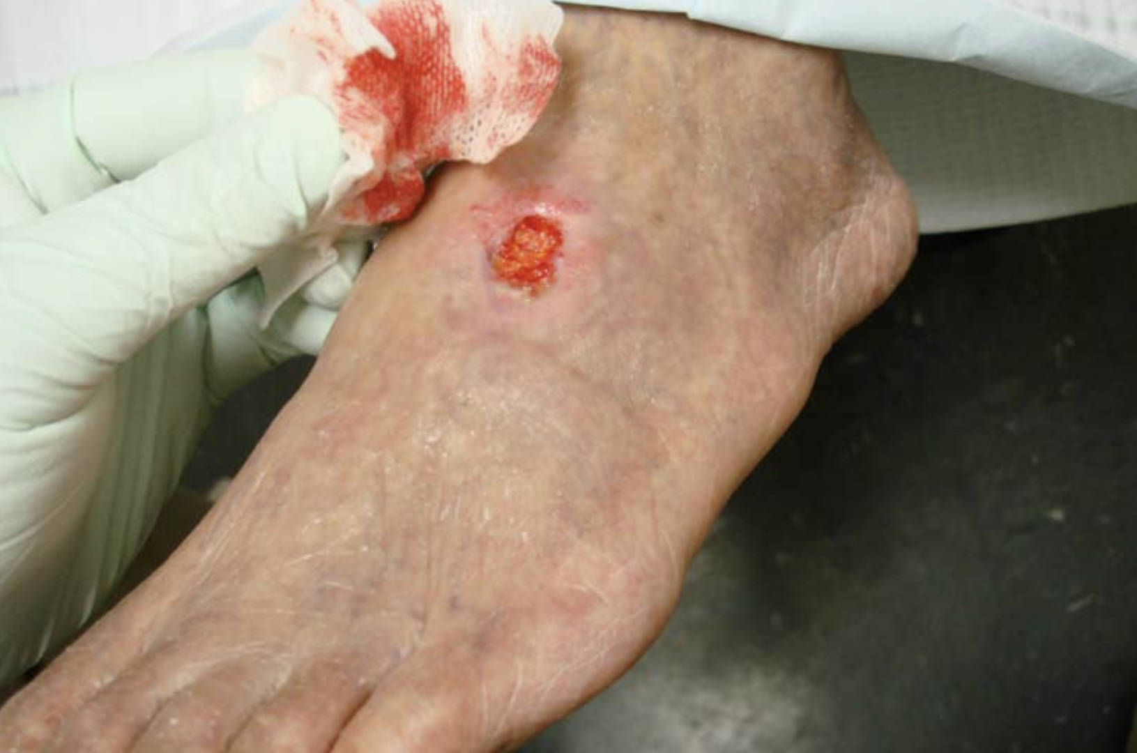
The image displayed illustrates a severe clinical presentation of a non-healing dorsal foot ulcer in a 71-year-old male patient with a history of diabetes and smoking. This visual evidence highlights the critical nature of circulatory compromise, where a wound measuring 2.5 cm by 2.4 cm has remained chronically open for nearly two years due to insufficient blood flow. Understanding the pathophysiology behind such wounds is crucial for recognizing the advanced stages of vascular disease and the importance of multidisciplinary care in limb preservation.
Understanding the Clinical Presentation
The photograph reveals a classic arterial ulcer located on the dorsum (top) of the foot, a common site for trauma and pressure in patients with compromised circulation. Unlike venous ulcers, which are often wet and located on the lower leg, this wound appears relatively dry with well-defined margins, often described as having a “punched-out” appearance. The tissue within the ulcer bed lacks the robust, red granulation tissue typically seen in healthy healing wounds, indicating significant ischemia—a restriction in blood supply to the tissues.
This specific case underscores the “perfect storm” of risk factors: advanced age, diabetes mellitus, and a history of smoking. Peripheral arterial disease (PAD) acts as the primary driver for this chronicity. In PAD, the arteries supplying the lower extremities become narrowed or blocked by plaque, preventing the delivery of oxygen and nutrients essential for tissue repair. When combined with diabetes, which can cause nerve damage (neuropathy) and microvascular dysfunction, the skin becomes fragile and unable to heal from even minor injuries.
The persistence of this ulcer for two years suggests that the patient likely suffers from critical limb ischemia, the most severe form of PAD. Without adequate blood flow, the body’s natural immune response and cellular repair mechanisms are halted. Clinicians evaluating such wounds look for specific signs that differentiate arterial ulcers from other wound types:
- Location: Typically on the toes, heels, or dorsum of the foot.
- Appearance: Deep, pale wound bed with well-defined edges; often covered in slough or necrotic tissue.
- Surrounding Skin: The skin may appear shiny, thin, and hairless due to poor perfusion.
- Pulse: Dorsalis pedis and posterior tibial pulses are usually weak or absent.
- Temperature: The affected foot is often cool to the touch compared to the contralateral limb.
The Pathophysiology of Peripheral Arterial Disease
Peripheral arterial disease is a systemic manifestation of atherosclerosis, a condition where fatty deposits, cholesterol, and calcium build up within the inner walls of the arteries. Over time, these plaques harden and narrow the vessel lumen, restricting blood flow. In the context of the lower extremities, this reduction in arterial flow means that muscles and skin do not receive adequate oxygen, especially during periods of increased demand, such as walking. In severe cases, the blood supply is insufficient even at rest, leading to the development of ischemic ulcers and gangrene.
Smoking is a potent accelerant of this process. Nicotine causes vasoconstriction (tightening of blood vessels) and damages the endothelial lining of the arteries, promoting plaque formation and clot development. For the patient in the image, long-term smoking likely caused widespread arterial damage, making the large vessels in the leg rigid and narrow. This systemic damage complicates treatment, as revascularization (restoring blood flow) becomes more difficult when multiple arterial segments are affected.
The Role of Diabetes in Chronic Ulceration
Diabetes mellitus complicates PAD through several mechanisms. High blood glucose levels damage both blood vessels and nerves over time. Diabetic neuropathy—the loss of sensation in the feet—is particularly dangerous. A patient may sustain a minor cut, blister, or pressure injury from tight shoes but feel no pain due to nerve damage. Consequently, they may continue to walk on the injury, worsening the wound.
Furthermore, diabetes affects the microcirculation (the tiny capillaries), thickening the basement membrane and impairing the exchange of nutrients and waste products. This creates a diabetic foot ulcer that is resistant to standard wound care. In the case described, the combination of macrovascular disease (PAD blocking large arteries) and microvascular dysfunction (diabetes affecting capillaries) results in a wound that remains stagnant for years. The immune system is also impaired in diabetic patients, increasing the risk of secondary bacterial infections that can spread to the bone (osteomyelitis), further threatening the limb.
Conclusion
The chronic dorsal foot ulcer depicted serves as a stark reminder of the debilitating effects of severe peripheral arterial disease, particularly when compounded by diabetes and smoking. These wounds are not merely skin deep; they are external markers of profound systemic vascular compromise. Successful management requires a comprehensive approach, including vascular assessment for potential surgical revascularization, strict glycemic control, smoking cessation, and advanced wound care therapies. Early recognition and aggressive intervention are vital to prevent amputation and improve the quality of life for patients suffering from these complex arterial conditions.

