The male external genitalia exhibits variations in its natural presentation, primarily influenced by the presence or absence of the foreskin. This diagram offers a clear comparative view of both an uncircumcised and a circumcised penis, highlighting their key external anatomical features. Understanding these distinct configurations is crucial for comprehensive anatomical knowledge, discussions on penile hygiene, and clinical considerations related to male reproductive health.
Dissecting the External Penile Anatomy
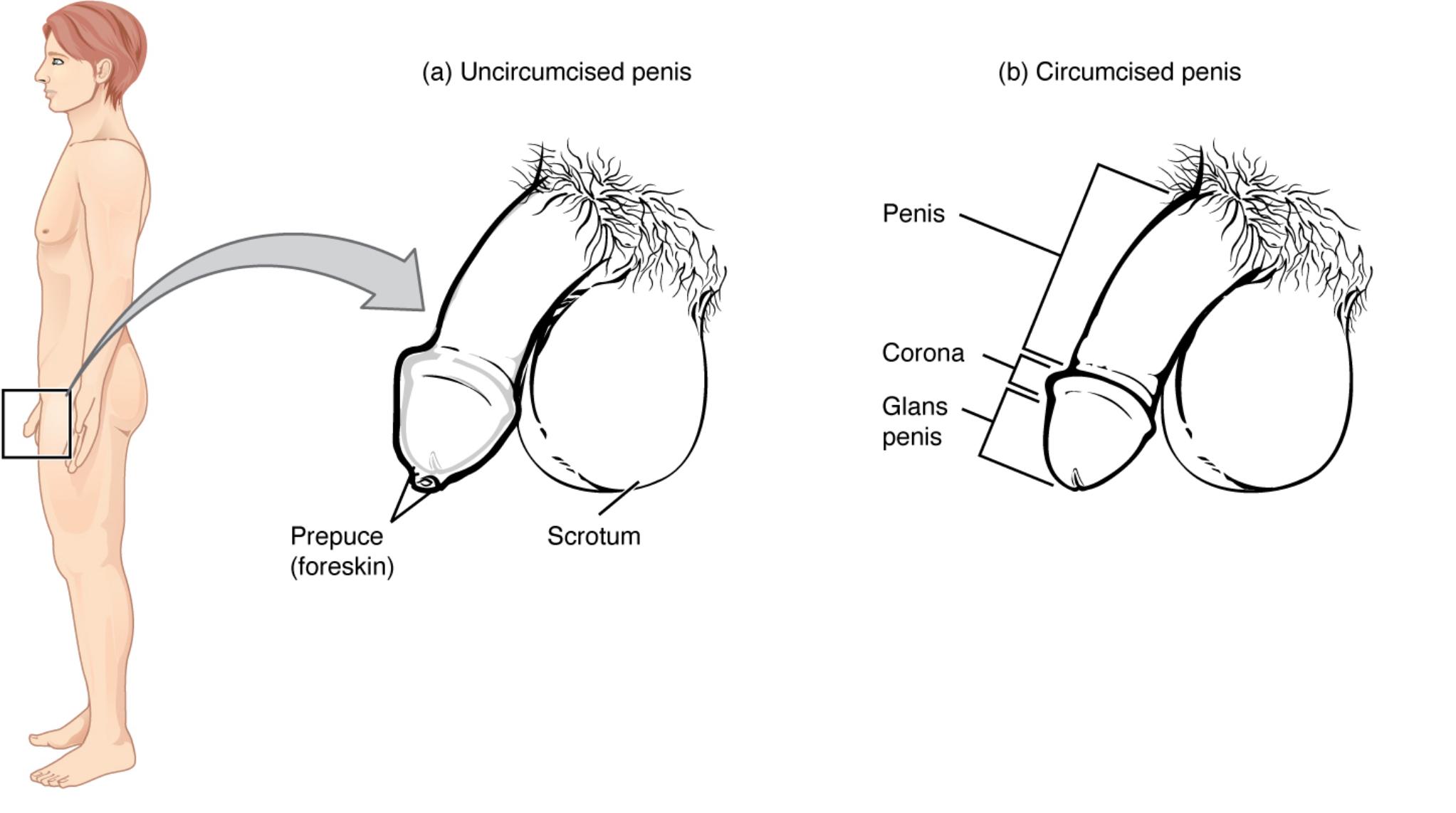
a) Uncircumcised penis: This illustration depicts the external appearance of an uncircumcised penis, characterized by the presence of a prepuce, or foreskin, covering the glans penis. This represents the natural anatomical state for males who have not undergone circumcision.
Prepuce (foreskin): A retractable fold of skin that naturally covers and protects the glans penis. In an uncircumcised individual, this skin can be moved back to expose the glans for cleaning or during sexual activity. Its presence is a defining characteristic of the uncircumcised penis.
Scrotum: A sac of skin and superficial fascia suspended inferior to the pubic symphysis and anterior to the anus. It contains the testes, epididymides, and the initial parts of the spermatic cords, maintaining a temperature suitable for spermatogenesis.
b) Circumcised penis: This illustration shows the external appearance of a circumcised penis, where the prepuce (foreskin) has been surgically removed, leaving the glans penis exposed. This is a common practice in many cultures and for various medical or religious reasons.
Penis: The primary external male copulatory organ, through which both urine and semen are expelled from the body. It consists of a shaft and the glans penis, and its internal structure involves erectile tissues like the corpora cavernosa and corpus spongiosum.
Corona: The crown-like ridge located at the base of the glans penis, separating the glans from the shaft. It is a highly sensitive area.
Glans penis: The enlarged, rounded tip of the penis, which is richly innervated and highly sensitive. In an uncircumcised penis, it is covered by the prepuce, while in a circumcised penis, it is exposed.
Understanding Penile Variations
This diagram provides a clear and essential comparative view of the uncircumcised and circumcised penis, illustrating the primary anatomical difference between the two: the presence or absence of the foreskin. The penis is the central external male organ, serving both urinary and reproductive functions. Its external morphology, particularly the glans and the surrounding structures, is crucial for both function and health.
In an uncircumcised penis (a), the glans is covered by the prepuce, or foreskin. This retractable fold of skin offers protection to the glans, helps maintain its moisture, and contributes to sensory experiences. Proper hygiene, which involves gentle retraction and cleaning, is important for uncircumcised individuals.
Conversely, in a circumcised penis (b), the foreskin has been surgically removed, leaving the glans penis permanently exposed. This altered anatomy reveals the glans and the corona, the ridge that separates the glans from the shaft. This anatomical variation is influenced by cultural, religious, and medical factors, and it also affects hygiene practices and potentially sensory perception.
Both anatomical presentations are normal variations in male anatomy, and understanding their specific features is important for healthcare professionals and individuals alike. It allows for informed discussions about hygiene, sexual health, and potential medical conditions that may arise in either state. The scrotum, depicted alongside the penis in both illustrations, is the external sac containing the testes, vital for sperm production.
The Penis: A Comparative Anatomical Study of Circumcised and Uncircumcised States
The human male external genitalia, particularly the penis, presents with anatomical variations that hold significant clinical, cultural, and personal relevance. This diagram offers a clear and side-by-side comparison of the uncircumcised and circumcised penis, effectively illustrating the defining anatomical distinctions between these two states. Such a visual aid is fundamental for a comprehensive understanding of male anatomy, hygiene practices, and the considerations surrounding the surgical procedure of circumcision.
In the uncircumcised penis (figure a), the glans penis, which is the sensitive, bulbous tip of the organ, is naturally covered by the prepuce, commonly known as the foreskin. This retractable fold of skin serves several physiological purposes: it protects the delicate glans from mechanical trauma, maintains its natural moisture, and contributes to sensory input during sexual activity through a rich network of nerve endings. For individuals with an uncircumcised penis, proper hygiene involves regular and gentle retraction of the foreskin to clean the glans and the sulcus (the groove behind the corona) to prevent the accumulation of smegma and reduce the risk of infection. Conditions such as phimosis (a tight foreskin that cannot be retracted) and paraphimosis (a retracted foreskin that cannot be returned to its normal position) are specific to the uncircumcised state and may necessitate medical intervention.
Conversely, the circumcised penis (figure b) is characterized by the surgical removal of the foreskin, leaving the glans penis permanently exposed. This anatomical configuration reveals the distinct corona, a ridge that separates the glans from the penile shaft. The practice of circumcision is deeply rooted in various cultural and religious traditions globally, and it is also performed for medical reasons, such as to treat or prevent recurrent balanitis (inflammation of the glans) or certain urinary tract infections. The constant exposure of the glans in a circumcised penis can lead to a slight keratinization or toughening of its surface over time. While the absence of the foreskin necessitates different hygiene routines, it is important to note that both uncircumcised and circumcised penile anatomies are considered normal variations.
Understanding these anatomical differences is crucial for healthcare professionals in providing appropriate care, for counseling individuals on hygiene practices, and for discussing the potential benefits and risks associated with circumcision. Both configurations contribute to the male reproductive and urinary functions, with the urethra traversing the length of the penis to allow for the passage of urine and semen. The scrotum, also depicted, plays the vital role of housing and regulating the temperature of the testes, which are responsible for sperm and hormone production.
Conclusion
This comparative diagram of the uncircumcised and circumcised penis offers a clear and concise visual explanation of their key external anatomical differences. By highlighting the presence or absence of the foreskin and other defining features like the glans and corona, it provides essential anatomical knowledge. This understanding is fundamental for informed discussions on male health, hygiene practices, and the diverse presentations of male external genitalia in both clinical and general contexts.

