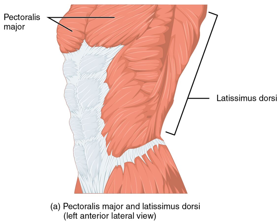
Understanding the anatomy of the human body is essential for appreciating how muscles contribute to movement and stability. The image provided showcases the Pectoralis major and Latissimus dorsi from a left anterior lateral view, highlighting their structure and positioning. This detailed visual aid serves as a valuable resource for exploring the muscular system’s complexity and its role in daily activities.
Label Introduction
Pectoralis major
The Pectoralis major is a large, fan-shaped muscle located in the upper chest region, playing a crucial role in shoulder movement. It facilitates actions such as flexion, adduction, and internal rotation of the humerus, making it vital for pushing and lifting motions.
Latissimus dorsi
The Latissimus dorsi is a broad, flat muscle that spans the lower and mid-back, extending to the sides of the torso. It is responsible for extending, adducting, and internally rotating the arm, contributing significantly to pulling movements and overall upper body strength.
Multifidus
The Multifidus is a series of small muscles located along the vertebral column, extending from the sacrum to the cervical region. It plays a key role in stabilizing the spine and facilitating segmental movements, contributing to posture and spinal alignment.
Practical Applications in Exercise and Therapy
Incorporating the Pectoralis major and Latissimus dorsi into fitness routines can yield significant benefits. This part provides actionable advice for optimizing their use in exercise and therapeutic settings. Understanding their anatomy aids in designing effective workout plans.
- Recommends exercises like push-ups to strengthen the Pectoralis major.
- Suggests lat pulldowns to target the Latissimus dorsi effectively.
- Advises on stretching routines to maintain flexibility and prevent tightness.
- Encourages consultation with professionals for personalized training programs.
Deep Spinal Muscles Anatomy
Exploring the intricate anatomy of the human spine reveals the critical role of deep spinal muscles. The provided image illustrates the deep spinal muscles with the multifidus removed, offering a clear view of the underlying structures from a detailed perspective. This visual resource is invaluable for understanding spinal stability and the muscular support system.
Overview of Deep Spinal Muscles Anatomy
This section provides an in-depth look at the deep spinal muscles, with the multifidus removed to expose underlying structures. These muscles are essential for maintaining spinal integrity and supporting the body’s core. The image highlights the complexity of the spinal musculature, aiding in anatomical studies.
- Reveals the layered arrangement of deep spinal muscles post-multifidus removal.
- Shows the connection points along the vertebrae, enhancing spinal support.
- Illustrates the role of adjacent muscles in compensating for the multifidus‘ absence.
- Offers a clear view of the erector spinae and other deep muscle groups.
Functions and Importance in Spinal Stability
The multifidus and related deep spinal muscles are vital for spinal health. This area explains their physiological functions, emphasizing their contribution to stability. Their coordinated action helps prevent injuries and supports proper posture.
- Enables the multifidus to provide segmental stability between vertebrae.
- Supports the spine during dynamic movements like bending and twisting.
- Contributes to load distribution across the vertebral column.
- Highlights the muscle’s role in proprioception and balance.
Clinical Relevance and Muscle Health
The health of the multifidus is critical for avoiding spinal issues. This section addresses potential conditions and therapeutic approaches related to deep spinal muscles. Maintaining their strength is key to long-term spinal function.
- Discusses multifidus atrophy, often linked to chronic lower back pain.
- Notes the impact of removing the multifidus on spinal biomechanics.
- Suggests core strengthening exercises to support deep spinal muscles.
- Emphasizes the need for early intervention in spinal muscle injuries.
Practical Applications in Rehabilitation
Rehabilitating the multifidus and deep spinal muscles involves targeted strategies. This part offers guidance on exercises and therapies to restore function. Understanding their anatomy enhances recovery outcomes.
- Recommends planks and bird-dog exercises to engage deep spinal muscles.
- Suggests physical therapy to rebuild multifidus strength post-injury.
- Advises on posture correction to reduce strain on spinal muscles.
- Encourages regular assessment by healthcare professionals.
Conclusion
The deep spinal muscles, with the multifidus removed, provide a unique perspective on spinal anatomy and support. This image serves as an educational cornerstone, shedding light on the muscles’ roles in stability and movement. By studying these structures, one can better address spinal health, implement effective rehabilitation, and enhance overall physical resilience.

