The pectoral girdle muscles are crucial for stabilizing the shoulder complex, creating a reliable foundation for arm movements by supporting the clavicle and scapula. This detailed analysis of the pectoral girdle muscles in a left anterior lateral view, with the pectoralis major and deltoid cut away, reveals the deeper muscles
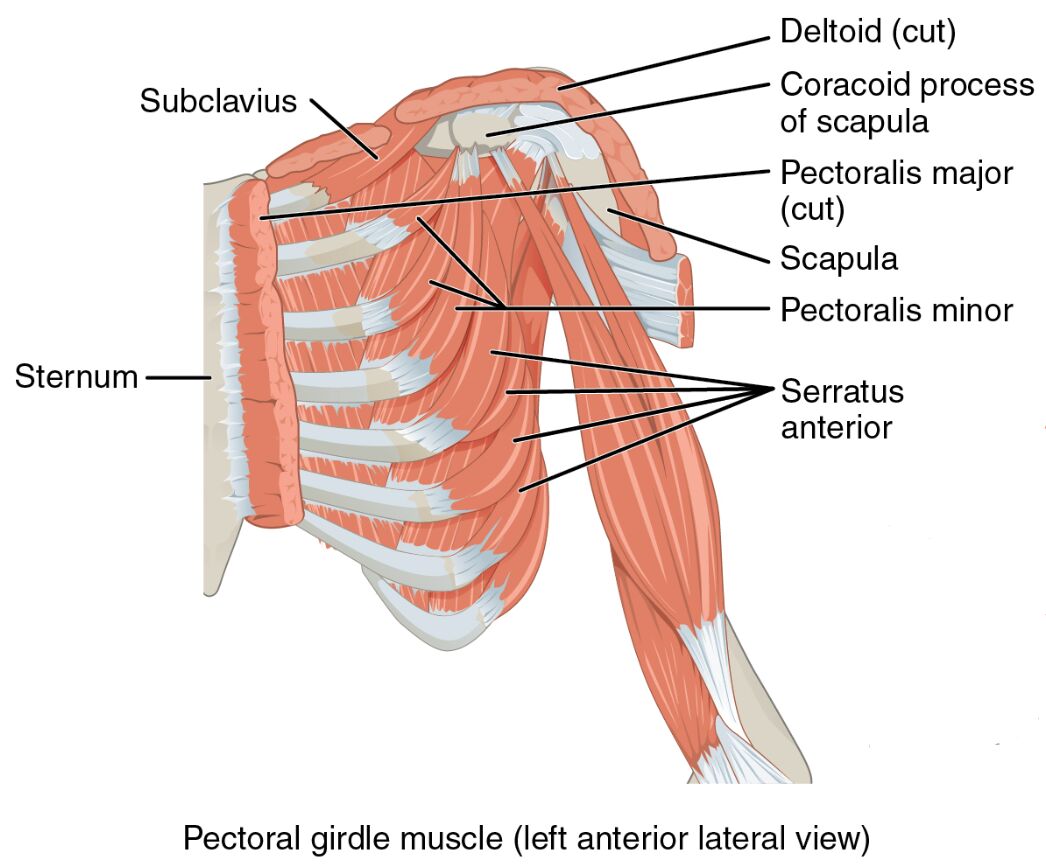
that position the girdle, offering essential insights into upper body anatomy.
Labeled Parts Introduction
Clavicle
The clavicle is a slender, S-shaped bone connecting the sternum to the scapula, serving as a critical attachment point for muscles that stabilize the pectoral girdle, and supports the weight of the upper limb. It enables smooth movement of the shoulder by maintaining its alignment.
Scapula
The scapula is a flat, triangular bone on the back, acting as the primary site for muscle attachments that position the pectoral girdle, and facilitates a wide range of shoulder motions. Its mobility is enhanced by the coordinated action of surrounding muscles.
Serratus anterior
The serratus anterior is a fan-shaped muscle along the lateral chest, originating from the ribs and inserting into the scapula, and protracts the scapula while stabilizing the pectoral girdle. It prevents winging of the scapula and supports pushing actions.
Pectoralis minor
The pectoralis minor is a thin muscle beneath the pectoralis major, attaching from the ribs to the scapula, and depresses the scapula while aiding in pectoral girdle stability. It also assists in elevating the ribs during deep breathing.
Subclavius
The subclavius is a small muscle under the clavicle, connecting it to the first rib, and stabilizes the clavicle while protecting underlying neurovascular structures. It minimizes excessive clavicular movement during arm elevation.
Trapezius
The trapezius is a large, diamond-shaped muscle covering the upper back and neck, attaching to the clavicle and scapula, and elevates, retracts, and rotates the scapula to position the pectoral girdle. It is vital for posture and upper limb coordination.
Levator scapulae
The levator scapulae is a narrow muscle running from the cervical vertebrae to the scapula, elevating the scapula and assisting in neck extension, and contributes to pectoral girdle stability. It supports shoulder height during arm-raising activities.
Rhomboid major
The rhomboid major is a broad muscle beneath the trapezius, connecting the spinal column to the scapula, and retracts and stabilizes the scapula to support the pectoral girdle. It works with the rhomboid minor to maintain scapular position.
Rhomboid minor
The rhomboid minor is a smaller muscle above the rhomboid major, linking the cervical and thoracic vertebrae to the scapula, and retracts the scapula while aiding in its stabilization. It enhances the coordinated function of the pectoral girdle.
Overview of Pectoral Girdle Muscle Anatomy
The pectoral girdle muscles provide a stable base for arm movement, with the clavicle and scapula serving as key anchors in this left anterior lateral view. The serratus anterior, trapezius, and other deeper muscles are exposed with the pectoralis major and deltoid cut away, highlighting their role in shoulder stability. This arrangement ensures effective upper body function and mobility.
- Forms a dynamic platform that supports the humerus during various motions.
- Protects the shoulder joint by maintaining proper structural alignment.
Structure of Pectoral Girdle Positioning Muscles
The trapezius and levator scapulae extend from the spine to the scapula, while the rhomboid major and rhomboid minor reinforce scapular retraction. The pectoralis minor and subclavius attach to the clavicle, providing additional stability, with the serratus anterior anchoring the scapula to the ribs.
- The trapezius offers extensive coverage for diverse scapular adjustments.
- The serratus anterior provides robust lateral support to the pectoral girdle.
Role in Shoulder Stability and Movement
The serratus anterior protracts the scapula during pushing movements, while the trapezius and rhomboid major retract it for pulling actions. The levator scapulae and pectoralis minor adjust scapular elevation and depression, with the subclavius stabilizing the clavicle during arm raises.
- The rhomboid minor supports scapular adduction for upright posture.
- The subclavius reduces clavicular strain during overhead motions.
Clinical Relevance and Physical Health
The serratus anterior is often weakened in scapular winging, necessitating targeted exercises, while trapezius tightness can lead to neck discomfort. The pectoralis minor may contribute to shoulder impingement if overly tight, affecting pectoral girdle function.
- A strained rhomboid major can restrict scapular retraction.
- The clavicle is a key indicator for diagnosing shoulder injuries.
Physical Examination and Rehabilitation
Physical exams assess the scapula for mobility and the clavicle for alignment, with the trapezius evaluated for tension. Rehabilitation focuses on strengthening the serratus anterior and stretching the levator scapulae to improve shoulder mechanics.
- Proper scapula movement enhances upper limb functionality.
- The pectoralis minor is addressed to alleviate shoulder tightness.
Conclusion
The pectoral girdle muscles, as illustrated in this left anterior lateral view with the pectoralis major and deltoid dissected, form a resilient system that stabilizes the clavicle and scapula for arm movement. From the serratus anterior to the rhomboid minor, these muscles ensure shoulder integrity and support. A thorough understanding of their anatomy aids in diagnosing and treating upper body conditions effectively.

