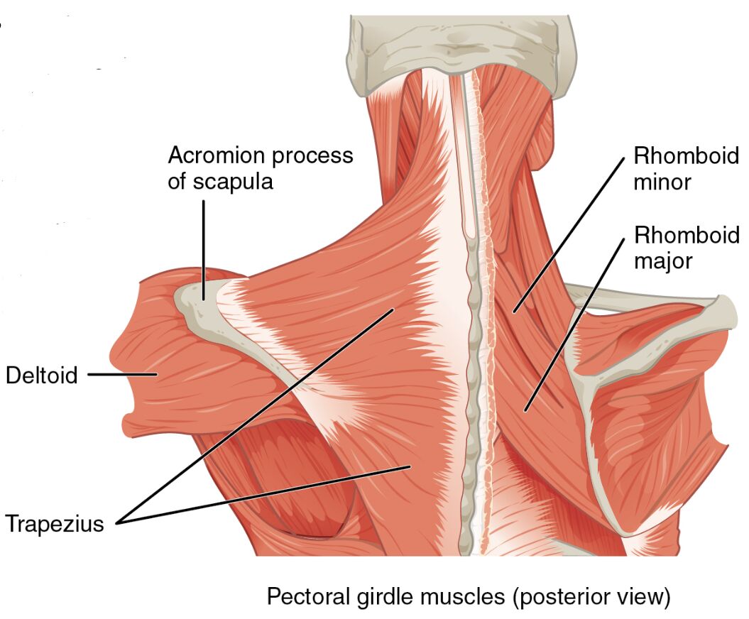
The pectoral girdle muscles are vital for stabilizing the shoulder complex, offering a solid foundation for arm movements by anchoring the scapula and clavicle. This detailed examination of the pectoral girdle muscles in a posterior view, with the pectoralis major and deltoid cut away, reveals the deeper muscles responsible for positioning the girdle, providing key insights into upper body anatomy.
Labeled Parts Introduction
Clavicle
The clavicle is a slender, S-shaped bone linking the sternum to the scapula, serving as an upper attachment for muscles that stabilize the pectoral girdle, and supports the weight of the upper limb. It maintains the shoulder’s alignment during movement.
Scapula
The scapula is a flat, triangular bone on the back, acting as the central attachment site for muscles that position the pectoral girdle, and enables a wide range of shoulder motions. Its mobility is controlled by the surrounding musculature.
Trapezius
The trapezius is a large, diamond-shaped muscle covering the upper back and neck, attaching to the clavicle and scapula, and elevates, retracts, and rotates the scapula to stabilize the pectoral girdle. It is essential for maintaining posture and coordinating arm movements.
Levator scapulae
The levator scapulae is a narrow muscle running from the cervical vertebrae to the scapula, elevating the scapula and assisting in neck extension, and contributes to pectoral girdle stability. It supports shoulder elevation during arm-raising tasks.
Rhomboid major
The rhomboid major is a broad muscle beneath the trapezius, connecting the spinal column to the scapula, and retracts and stabilizes the scapula to support the pectoral girdle. It works with the rhomboid minor to hold the scapula in place.
Rhomboid minor
The rhomboid minor is a smaller muscle above the rhomboid major, linking the cervical and thoracic vertebrae to the scapula, and retracts the scapula while aiding in its stabilization. It enhances the coordinated positioning of the pectoral girdle.
Spine of scapula
The spine of scapula is a prominent ridge on the posterior scapula, serving as an attachment and landmark for muscles like the trapezius, and provides structural support to the shoulder blade. It helps guide muscle forces during scapular movement.
Infraspinatus
The infraspinatus is a thick muscle covering the posterior scapula below the spine, rotating the humerus externally and stabilizing the shoulder joint, and supports the pectoral girdle indirectly. It assists in maintaining arm position during motion.
Teres minor
The teres minor is a narrow muscle below the infraspinatus, attaching from the scapula to the humerus, and externally rotates the arm while stabilizing the shoulder. It contributes to the overall stability of the pectoral girdle during arm movements.
Supraspinatus
The supraspinatus is a muscle above the spine of scapula, originating from the scapula and inserting into the humerus, and initiates arm abduction while stabilizing the shoulder joint. It plays a supportive role in pectoral girdle function.
Overview of Pectoral Girdle Muscle Anatomy
The pectoral girdle muscles provide a stable platform for arm movement, with the clavicle and scapula serving as primary anchors in this posterior view. The trapezius, rhomboid major, and other deeper muscles are exposed with the pectoralis major and deltoid cut away, highlighting their role in shoulder stability. This muscular network ensures effective upper body mechanics.
- Establishes a firm base for the humerus to pivot during lifting or pulling.
- Safeguards the shoulder joint by maintaining proper alignment.
Structure of Pectoral Girdle Positioning Muscles
The trapezius and levator scapulae extend from the spine to the scapula, while the rhomboid major and rhomboid minor reinforce scapular retraction. The spine of scapula acts as a structural ridge, supporting attachments for the infraspinatus, teres minor, and supraspinatus, which stabilize the shoulder.
- The trapezius provides extensive coverage for scapular adjustments.
- The spine of scapula serves as a critical landmark for muscle orientation.
Role in Shoulder Stability and Movement
The trapezius elevates and retracts the scapula for pulling motions, while the rhomboid major and rhomboid minor enhance retraction for posture. The levator scapulae adjusts scapular height, and the infraspinatus, teres minor, and supraspinatus stabilize the humerus, supporting pectoral girdle function.
- The rhomboid minor aids in scapular adduction for upright stance.
- The supraspinatus initiates abduction to support arm raising.
Clinical Relevance and Physical Health
The trapezius can become tight, leading to neck pain, while rhomboid major strain may limit scapular retraction. The infraspinatus and teres minor are often assessed for rotator cuff issues, which can affect pectoral girdle stability.
- Weakness in the levator scapulae may cause shoulder drooping.
- The scapula is a key focus for diagnosing winging or misalignment.
Physical Examination and Rehabilitation
Physical exams evaluate the clavicle for alignment and the scapula for mobility, with the trapezius checked for tension. Rehabilitation targets strengthening the rhomboid minor and stretching the levator scapulae to improve shoulder function.
- Proper scapula positioning enhances upper limb performance.
- The infraspinatus is strengthened to support rotator cuff health.
Conclusion
The pectoral girdle muscles, as depicted in this posterior view with the pectoralis major and deltoid dissected, form a robust system that stabilizes the clavicle and scapula for arm movement. From the trapezius to the supraspinatus, these muscles ensure shoulder integrity and support. A deep understanding of their anatomy aids in diagnosing and treating upper body conditions effectively.

