The pectoral girdle, a critical component of the upper body, consists of the clavicle and scapula, which connect the upper limb to the axial skeleton at the sternum. This anatomical structure provides stability and mobility, enabling a wide range of arm movements essential for daily activities. Understanding its components and their functions is vital for professionals in anatomy, orthopedics, and physical therapy. This article explores the detailed anatomy of the pectoral girdle, including its labeled parts and their roles, offering a comprehensive guide to its structure and significance.
Labeled Parts of the Pectoral Girdle
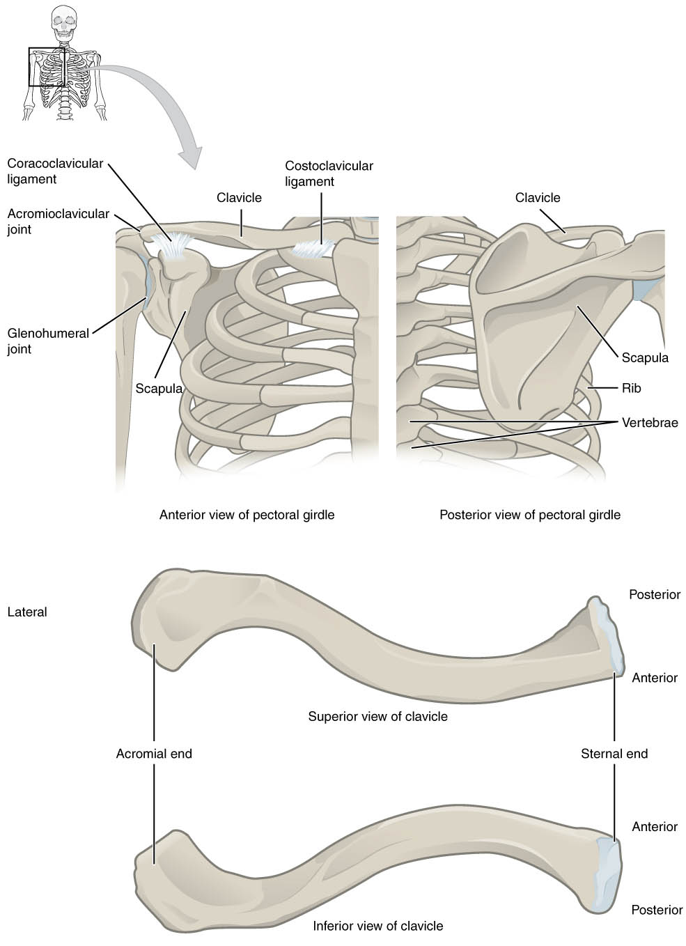
Clavicle
The clavicle, commonly known as the collarbone, is a long, S-shaped bone that forms the anterior part of the pectoral girdle. It connects the sternum to the scapula, providing structural support and serving as a strut to keep the shoulder in place.
Scapula
The scapula, or shoulder blade, is a flat, triangular bone located on the posterior side of the pectoral girdle. It articulates with the clavicle and humerus, facilitating shoulder movement and housing the glenoid cavity for the glenohumeral joint.
Coracoclavicular Ligament
This ligament connects the clavicle to the coracoid process of the scapula, providing stability to the acromioclavicular joint. It plays a key role in preventing excessive movement and maintaining proper alignment during shoulder motion.
Acromioclavicular Joint
The acromioclavicular joint is where the clavicle meets the acromion of the scapula, allowing limited movement while providing stability. It is crucial for shoulder elevation and rotation, often involved in injuries like shoulder separations.
Glenohumeral Joint
The glenohumeral joint, or shoulder joint, is a ball-and-socket joint formed by the head of the humerus and the glenoid cavity of the scapula. It allows a wide range of motion, including flexion, extension, abduction, and rotation of the arm.
Costoclavicular Ligament
The costoclavicular ligament anchors the clavicle to the first rib, stabilizing the sternoclavicular joint. It helps limit excessive upward movement of the clavicle during shoulder motion.
Ribs
The ribs are curved bones that form the thoracic cage, with the first rib connecting to the clavicle via the costoclavicular ligament. They protect vital organs and support the pectoral girdle’s attachment to the axial skeleton.
Vertebrae
The vertebrae of the thoracic spine provide a posterior anchor for the scapula, which glides over them during shoulder movement. They contribute to the structural integrity of the upper back and support posture.
Acromial End
The acromial end of the clavicle is its lateral end, articulating with the acromion of the scapula at the acromioclavicular joint. This end is broader and flatter, facilitating joint stability.
Sternal End
The sternal end of the clavicle is its medial end, connecting to the sternum at the sternoclavicular joint. It is rounded and forms a synovial joint that allows slight movement during shoulder actions.
Anatomy of the Pectoral Girdle
Structure and Components
The pectoral girdle is a foundational structure for upper limb function. Its primary components, the clavicle and scapula, work together to ensure stability and mobility.
- The clavicle acts as a bridge between the axial skeleton and the upper limb, transmitting forces from the arm to the trunk.
- The scapula provides a broad surface for muscle attachments, such as the rotator cuff muscles, which stabilize the glenohumeral joint.
- Ligaments like the coracoclavicular and costoclavicular play a critical role in maintaining joint integrity during dynamic movements.
- The glenohumeral joint’s design as a ball-and-socket joint allows for the greatest range of motion in the body, making it essential for activities like throwing or lifting.
- The acromioclavicular joint, though smaller, supports shoulder elevation and is often a site of injury in athletes due to its limited range of motion.
Functional Role in Movement
The pectoral girdle facilitates a wide range of upper limb movements. Its structure ensures both strength and flexibility for daily tasks.
- The clavicle’s S-shape allows it to absorb and distribute mechanical forces, protecting the underlying neurovascular structures.
- The scapula’s ability to glide over the thoracic cage enables shoulder protraction, retraction, and rotation, essential for reaching and lifting.
- The glenohumeral joint’s mobility is balanced by the stability provided by ligaments and the rotator cuff muscles, preventing dislocations.
- The acromioclavicular joint and its ligaments work together to maintain shoulder alignment during overhead movements.
- The costoclavicular ligament ensures the clavicle remains anchored to the rib cage, preventing excessive elevation that could strain surrounding structures.
Clinical Significance
The pectoral girdle’s anatomy is crucial for diagnosing and treating shoulder-related conditions. Its components are often involved in injuries and degenerative conditions.
- Clavicle fractures are common due to its superficial location, often resulting from falls or direct trauma.
- Acromioclavicular joint injuries, such as separations, occur frequently in contact sports, requiring careful rehabilitation to restore function.
- The glenohumeral joint is prone to dislocations due to its wide range of motion and relatively shallow socket.
- Scapular dyskinesis, or abnormal scapular movement, can lead to shoulder impingement and pain, often treated with physical therapy.
- Ligament injuries, such as tears in the coracoclavicular ligament, may require surgical intervention to restore stability.
Physical Characteristics of the Pectoral Girdle
Bone Structure and Shape
The bones of the pectoral girdle are uniquely shaped to support their functions. Their design reflects a balance between strength and mobility.
- The clavicle is a long bone with a double curvature, making it resilient yet lightweight for its role in shoulder support.
- The scapula’s triangular shape provides a large surface for muscle attachments while minimizing weight.
- The acromial end of the clavicle is broader to accommodate articulation with the scapula, while the sternal end is more rounded for the sternoclavicular joint.
- The scapula’s glenoid cavity is shallow, prioritizing mobility over stability, which is compensated by surrounding ligaments and muscles.
- The ribs and vertebrae provide a stable foundation for the pectoral girdle, ensuring proper alignment during movement.
Ligamentous Support
Ligaments are essential for maintaining the pectoral girdle’s stability. They connect bones and limit excessive motion.
- The coracoclavicular ligament consists of two parts, the trapezoid and conoid ligaments, which together prevent superior displacement of the clavicle.
- The costoclavicular ligament is a strong, fibrous band that anchors the clavicle to the first rib, resisting upward forces.
- The acromioclavicular joint is reinforced by its own ligament, which prevents separation during minor impacts.
- The glenohumeral joint relies on capsular ligaments and the rotator cuff for stability, as its bony structure offers little inherent support.
- These ligaments collectively ensure the pectoral girdle can withstand the stresses of daily activities and athletic performance.
The pectoral girdle is a remarkable structure that bridges the upper limb to the axial skeleton, enabling a wide range of movements while providing stability. Its components, including the clavicle, scapula, and supporting ligaments, work in harmony to support daily activities and athletic endeavors. A thorough understanding of its anatomy and function is essential for diagnosing and managing shoulder-related conditions, ensuring optimal outcomes for patients with injuries or dysfunctions.

