The complex architecture of nerves is a testament to their critical role in coordinating bodily functions, supported by a series of connective tissue layers that provide both protection and organization. This article explores the labeled components of nerve structure, as depicted in a detailed diagram, offering insights into how these layers facilitate nerve impulse transmission and maintain neural integrity. Understanding this anatomy lays the foundation for appreciating the nerve’s resilience and its importance in overall health.
Labeled Structures in Nerve Anatomy
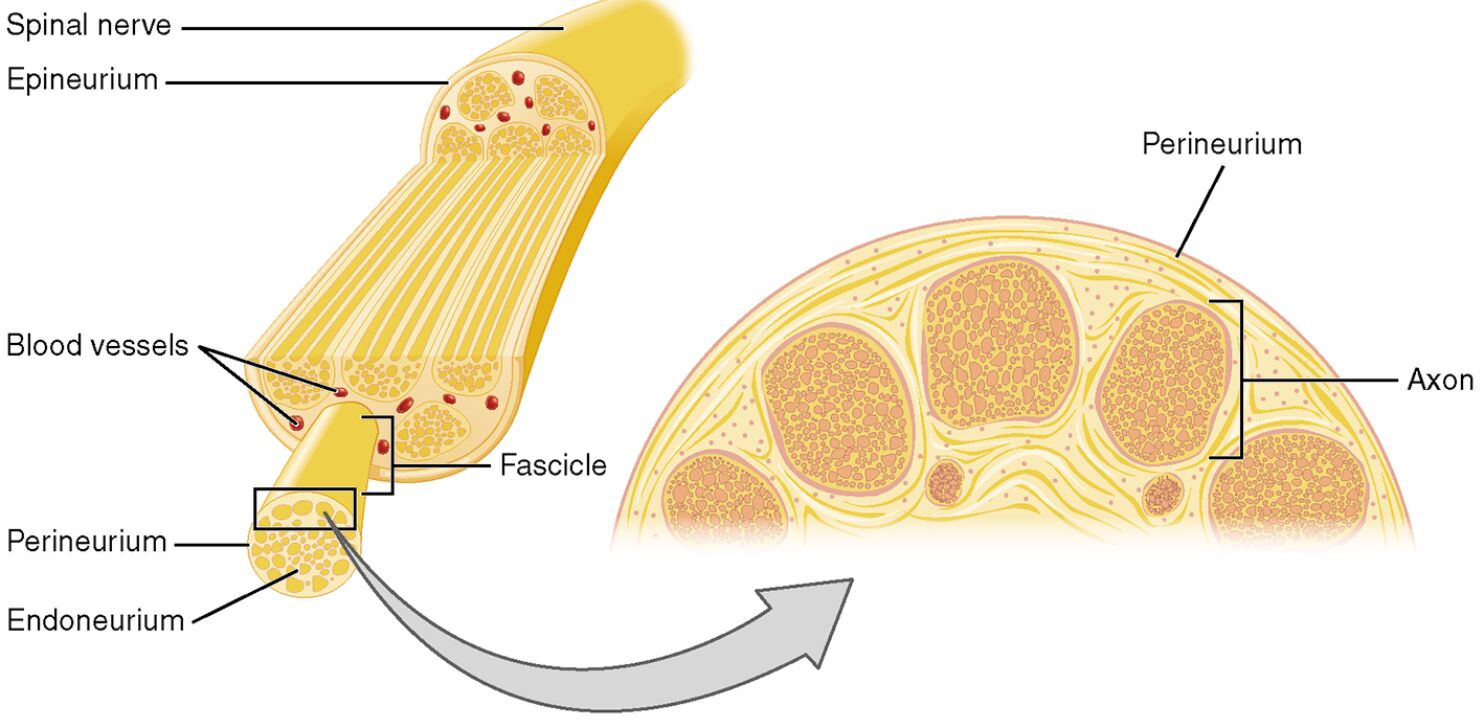
This section delves into each labeled component in the provided image, providing a comprehensive explanation of their roles and anatomical significance.
Spinal nerve The spinal nerve emerges from the spinal cord, serving as a mixed nerve that transmits motor, sensory, and autonomic signals to and from the body. It is encased by the epineurium, which bundles multiple fascicles into a cohesive unit for efficient signal conduction.
Epineurium The epineurium is the outermost layer of connective tissue surrounding the spinal nerve, offering protection against mechanical stress and external injury. It contains blood vessels and adipose tissue, ensuring a steady supply of nutrients and oxygen to the nerve’s internal structures.
Blood vessels The blood vessels embedded within the epineurium deliver oxygen and nutrients to the nerve tissue, supporting the metabolic demands of axons and surrounding cells. These vessels form a capillary network that extends into deeper layers, maintaining nerve health during activity.
Fascicle The fascicle is a bundle of nerve fibers grouped within the perineurium, organizing axons to enhance signal transmission efficiency. Multiple fascicles, held together by the epineurium, form the spinal nerve, allowing for specialized functional groupings.
Perineurium The perineurium is a multilayered connective tissue sheath encasing each fascicle, providing a protective barrier and maintaining a stable internal environment. It contributes to the blood-nerve barrier, safeguarding axons from potentially harmful substances.
Endoneurium The endoneurium is a delicate connective tissue layer surrounding individual axons within a fascicle, offering support and insulation for nerve fibers. It houses Schwann cells, which produce myelin to enhance the speed and efficiency of nerve impulses.
Axon The axon is the elongated projection of a neuron that conducts electrical impulses away from the cell body, forming the functional core of nerve fibers. In the diagram, axons are depicted within fascicles, surrounded by supportive connective tissue layers.
Anatomy of Nerve Structure
The layered organization of nerves ensures both structural stability and functional efficiency. This intricate design supports the nerve’s ability to transmit signals across the body.
- The epineurium, composed of dense collagen, encases the entire nerve, providing flexibility and resistance to stretching or compression.
- Fascicles within the nerve are compartmentalized by the perineurium, which also regulates the microenvironment to protect axonal function.
- The endoneurium supports individual axons, with Schwann cells in the peripheral nervous system producing myelin sheaths for rapid conduction.
- Blood vessels within the epineurium form a network that supplies oxygen, with a capillary bed extending into fascicles to meet metabolic needs.
Physiological Role in Nerve Function
The connective tissue layers of nerves play a crucial role in facilitating signal transmission and maintaining neural health. Their physiological contributions are essential for coordinated movement and sensory processing.
- The perineurium’s blood-nerve barrier controls the exchange of ions and molecules, protecting axons from inflammatory or toxic damage.
- Myelin, produced by Schwann cells in the endoneurium, enables saltatory conduction, significantly increasing the speed of nerve impulses.
- The epineurium’s vascular supply ensures a constant oxygen level, supporting the high metabolic rate of neurons, which rely on aerobic respiration.
- Fascicles group fibers with similar functions, such as motor or sensory, optimizing the nerve’s efficiency in processing and transmitting signals.
Clinical Significance and Microscopic Insights
The labeled nerve structure provides valuable insights into its organization and potential clinical implications. Understanding these layers aids in diagnosing and managing nerve-related conditions.
- Peripheral nerve injuries, such as those affecting the epineurium, can lead to neuromas if regeneration is disrupted, often requiring surgical repair.
- The perineurium’s barrier function is critical in conditions like diabetic neuropathy, where its compromise can result in axonal degeneration.
- The diagram, based on a 40x magnification micrograph, highlights fascicle arrangement, assisting in the identification of nerve compression or damage.
- Imaging modalities like ultrasound or MRI can detect epineurial thickening, guiding treatments for disorders such as carpal tunnel syndrome.
Nerve Regeneration and Connective Tissue Support
The connective tissue layers support the nerve’s regenerative capacity, a key factor in recovery following injury. This process underscores the nerve’s ability to adapt and heal.
- The epineurium provides a structural scaffold for regenerating axons, with blood vessels supplying essential nutrients during the repair phase.
- The perineurium forms bands of Büngner, guiding Schwann cells to direct axonal growth within fascicles after injury.
- The endoneurium’s Schwann cells secrete growth factors like nerve growth factor (NGF), promoting axon elongation and remyelination.
- Successful regeneration hinges on minimizing scar tissue within the epineurium, which can impede axonal regrowth if excessive.
In conclusion, the layered structure of nerves, from the protective epineurium to the supportive endoneurium, reflects a sophisticated system designed for signal transmission and resilience. The detailed anatomical insights provided by the labeled diagram enhance our understanding of nerve function, offering a solid basis for advancing diagnostic and therapeutic strategies in neurology.

