The intricate architecture of nerves is a marvel of biological engineering, supported by layers of connective tissue that ensure both protection and functionality. This article delves into the structural organization of nerves, as illustrated by a detailed diagram and microscopic view, highlighting the roles of the epineurium, perineurium, and endoneurium. Understanding these components provides valuable insights into nerve physiology and their clinical relevance in maintaining neural communication.
Labeled Structures in Nerve Anatomy
This section explores each labeled component in the provided images, offering a detailed explanation of their roles and anatomical significance.
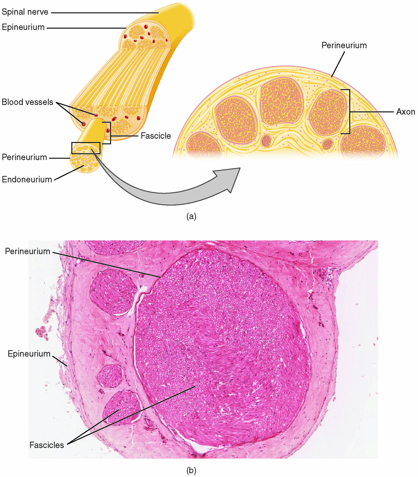
Spinal nerve The spinal nerve is a mixed nerve that emerges from the spinal cord, carrying both sensory and motor fibers to connect the central nervous system with the periphery. It is encased by the epineurium, which bundles multiple fascicles to form a functional unit for signal transmission.
Epineurium The epineurium is the outermost connective tissue layer surrounding the entire spinal nerve, providing structural support and protection against mechanical injury. It contains blood vessels and fat, cushioning the nerve and facilitating nutrient delivery to inner layers.
Blood vessels The blood vessels within the epineurium supply oxygen and nutrients to the nerve tissue, ensuring the metabolic needs of axons and supporting cells are met. They form a network that penetrates deeper layers, maintaining the nerve’s viability during activity.
Fascicle The fascicle is a bundle of nerve fibers grouped within the perineurium, organized to optimize signal conduction and protect individual axons. Multiple fascicles, held together by the epineurium, constitute the spinal nerve’s cross-sectional structure.
Perineurium The perineurium is a multilayered sheath of connective tissue encasing each fascicle, creating a protective barrier that regulates the internal environment. It also contributes to the blood-nerve barrier, preventing harmful substances from reaching the axons.
Endoneurium The endoneurium is a delicate layer of connective tissue surrounding individual axons within a fascicle, providing insulation and support for nerve fibers. It houses Schwann cells, which myelinate peripheral nerves, enhancing signal speed and efficiency.
Axon The axon is the elongated projection of a neuron that conducts electrical impulses away from the cell body, forming the core of nerve fiber function. In the microscopic view, axons are visible as the central structures within each fascicle, surrounded by supportive layers.
Anatomy of Nerve Structure
The nerve’s layered architecture ensures both structural integrity and functional efficiency. This organization is critical for the transmission of nerve impulses across the body.
- The epineurium, a dense collagenous layer, encases the entire nerve, offering flexibility and resistance to stretching.
- Fascicles within the nerve are compartmentalized by the perineurium, which also maintains a controlled microenvironment for axons.
- The endoneurium supports individual axons, with Schwann cells providing myelin sheaths in the peripheral nervous system to insulate and speed up conduction.
- Blood vessels embedded in the epineurium distribute nutrients, with a capillary network extending into the fascicles to support metabolic activity.
Physiological Role in Nerve Function
Nerve layers play a vital role in facilitating signal transmission and maintaining nerve health. Their physiological contributions are essential for coordinated movement and sensory perception.
- The perineurium’s blood-nerve barrier regulates ion and molecule exchange, protecting axons from inflammatory or toxic insults.
- Myelin, produced by Schwann cells in the endoneurium, increases conduction velocity by allowing saltatory conduction along the axon.
- The epineurium’s vascular supply ensures a constant oxygen level, supporting the high metabolic demand of nerve cells, which rely heavily on aerobic respiration.
- Fascicles group fibers with similar functions, such as motor or sensory, optimizing the nerve’s efficiency in signal processing.
Clinical Significance and Microscopic Insights
The microscopic view of nerve structure provides a window into its organization and potential clinical implications. Understanding these layers aids in diagnosing and treating nerve-related conditions.
- Peripheral nerve injuries, such as those affecting the epineurium, can lead to neuromas if regeneration is impaired, requiring surgical intervention.
- The perineurium’s integrity is crucial in conditions like diabetic neuropathy, where its barrier function may be compromised, leading to axonal damage.
- Histological examination at 40x magnification, as shown, reveals fascicle arrangement, aiding in the diagnosis of nerve compression or degeneration.
- Imaging techniques like ultrasound or MRI can assess epineurial thickening, guiding treatments for conditions like carpal tunnel syndrome.
Nerve Regeneration and Connective Tissue Support
The connective tissue layers support nerve regeneration, a process critical for recovery after injury. This regenerative capacity highlights the nerve’s adaptive potential.
- The epineurium guides regenerating axons by providing a structural scaffold, with blood vessels supplying necessary nutrients during repair.
- The perineurium forms bands of Büngner, aligning Schwann cells to direct axonal growth within fascicles.
- The endoneurium’s Schwann cells produce growth factors like nerve growth factor (NGF), promoting axon elongation and remyelination.
- Successful regeneration depends on minimizing scar tissue formation within the epineurium, which can obstruct axonal regrowth.
In conclusion, the layered structure of nerves, from the protective epineurium to the supportive endoneurium, exemplifies a sophisticated system designed for signal transmission and resilience. The microscopic insights offered by histological sections enhance our understanding of nerve function, paving the way for improved diagnostic and therapeutic approaches in neurology.

