The transition from fetal to neonatal circulation is one of the most remarkable physiological adaptations an individual undergoes. This intricate process involves significant restructuring of the cardiovascular system to accommodate independent respiratory and metabolic functions. This image provides a detailed visual guide to these critical changes, illustrating the key differences in blood flow pathways before and immediately after birth. Understanding these anatomical and functional shifts is fundamental to comprehending the unique aspects of newborn physiology and potential congenital anomalies.
Key Circulatory Adaptations Explained
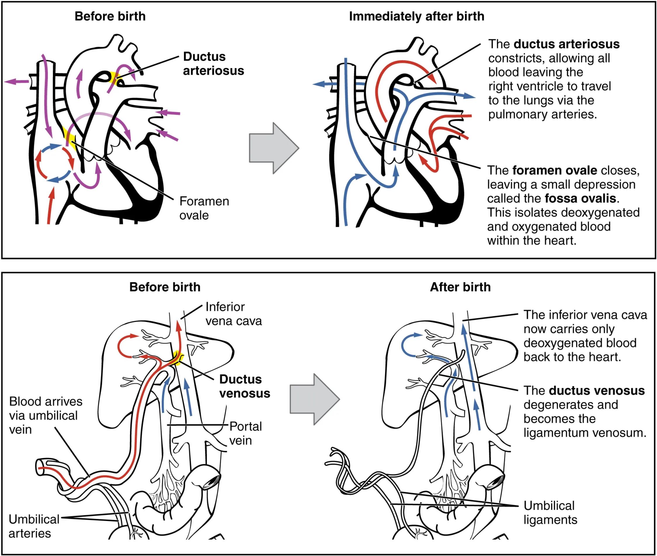
Ductus arteriosus: Before birth, this vital blood vessel connects the pulmonary artery to the aorta, allowing most of the blood from the right ventricle to bypass the non-functional fetal lungs. Immediately after birth, it constricts, diverting blood to the lungs for oxygenation and typically closes completely within days to weeks.
Foramen ovale: This is an opening in the septum between the right and left atria in the fetal heart, enabling oxygenated blood from the placenta (via the right atrium) to bypass the lungs and enter the systemic circulation directly. Post-birth, increased pressure in the left atrium compared to the right causes this flap-like opening to close, eventually forming the fossa ovalis.
Fossa ovalis: This is the anatomical remnant of the foramen ovale, a small depression in the interatrial septum of the heart. Its formation signifies the successful closure of the fetal shunt, ensuring complete separation of oxygenated and deoxygenated blood within the heart.
Inferior vena cava: In the fetal circulation, the inferior vena cava carries a mixture of highly oxygenated blood from the umbilical vein and deoxygenated blood from the lower body. After birth, with the cessation of placental circulation, it primarily carries only deoxygenated blood from the lower extremities and abdomen back to the heart.
Ductus venosus: This fetal shunt bypasses the liver, allowing oxygenated blood from the umbilical vein to directly enter the inferior vena cava and thus the systemic circulation. After birth, it degenerates and fibroses, becoming the ligamentum venosum.
Portal vein: In both fetal and adult circulation, the portal vein carries nutrient-rich blood from the gastrointestinal tract to the liver. In the fetus, some umbilical blood also flows into the portal system before reaching the inferior vena cava via the ductus venosus.
Umbilical vein: This single large vessel carries oxygenated, nutrient-rich blood from the placenta to the fetal liver and then, via the ductus venosus, to the inferior vena cava. After birth, with the cutting of the umbilical cord, the umbilical vein obliterates and becomes the ligamentum teres hepatis.
Umbilical arteries: These two arteries carry deoxygenated blood and waste products from the fetal internal iliac arteries back to the placenta. After birth, they constrict and largely obliterate, forming the medial umbilical ligaments.
Understanding the Transition
The neonatal circulatory system undergoes a profound transformation at birth, driven primarily by the initiation of breathing and the clamping of the umbilical cord. Before birth, the fetal lungs are fluid-filled and non-functional for gas exchange, and the placenta serves as the primary organ for nutrient and gas exchange. Consequently, the fetal circulation employs several shunts to bypass the lungs and liver:
- Ductus arteriosus: Shunts blood from the pulmonary artery to the aorta.
- Foramen ovale: Allows blood to bypass the right ventricle and pulmonary circulation by flowing directly from the right atrium to the left atrium.
- Ductus venosus: Allows oxygenated blood from the umbilical vein to bypass the hepatic circulation and flow directly into the inferior vena cava.
Upon the first breath, pulmonary vascular resistance significantly drops, leading to increased blood flow to the lungs. Concurrently, the clamping of the umbilical cord eliminates the low-resistance placental circulation, increasing systemic vascular resistance. These pressure changes, along with increased oxygen tension, trigger the closure of the fetal shunts. The ductus arteriosus constricts, redirecting blood flow to the now-functional lungs. The foramen ovale closes, preventing the mixing of oxygenated and deoxygenated blood within the heart. Finally, the ductus venosus degenerates, ensuring all portal blood passes through the liver.
This intricate sequence of events is crucial for establishing the independent postnatal circulation. Disruptions in this transition can lead to various congenital heart conditions, such as patent ductus arteriosus (PDA) or patent foramen ovale (PFO), which are often managed clinically to prevent long-term complications. The precise timing and coordination of these physiological changes highlight the remarkable adaptability of the human cardiovascular system.
Conclusion
The detailed diagrams of the neonatal circulatory system before and after birth offer an invaluable resource for understanding the critical physiological adaptations that occur during this pivotal life stage. From the temporary shunts that characterize fetal life to their dramatic closure and transformation into adult structures, each component plays a vital role in ensuring a smooth transition to independent existence. A thorough grasp of these processes is essential for medical professionals to diagnose and manage any deviations from normal development, underscoring the delicate balance inherent in the human circulatory system.

